


Testicular cancer is the most common solid malignant tumor in men aged 15–35.Approximately 95% of cases are germ cell tumors; among these, seminomas account for 50%–60%, and non-seminomas account for 40%–50%.With a cure rate exceeding 95%, it is one of the most curable solid malignant tumors.
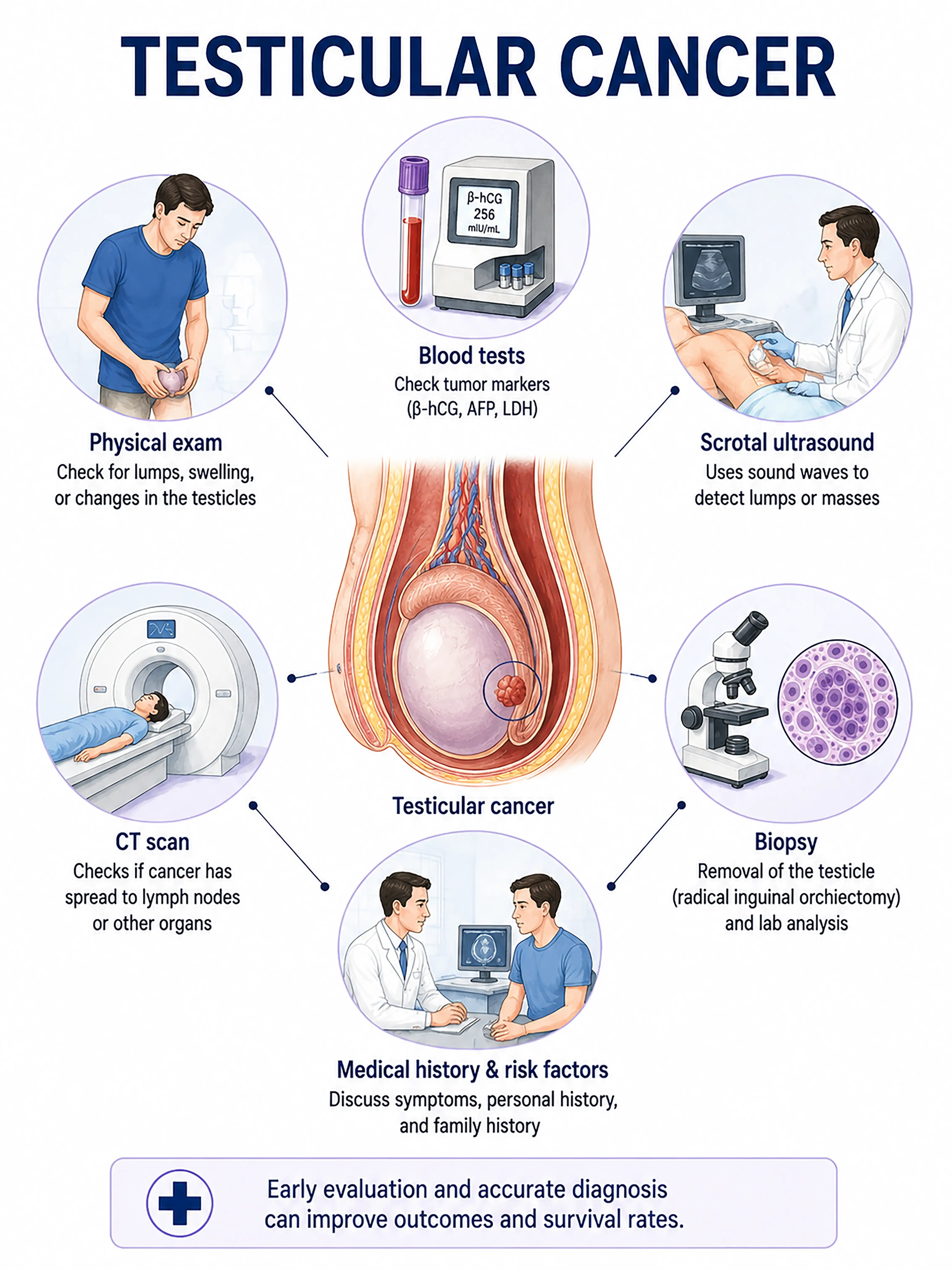
Diagnosis based on testicular cancer stage
1.Early diagnosis of testicular cancer
Painless testicular lump or enlargement.Scrotal ultrasound reveals a hypoechoic solid mass within the testis; serum AFP and β-hCG levels are abnormal; definitive diagnosis is made via orchiectomy and specimen analysis.
2.Diagnosis of intermediate-stage testicular cancer
Tumor enlargement accompanied by a sensation of heaviness.Ultrasound assesses tumor size and nature; CT evaluates retroperitoneal lymph node metastasis; tumor markers determine the stage (S1–S3).
3.Diagnosis of advanced testicular cancer
Distant metastasis to the lungs, liver, and brain.Whole-body CT and PET-CT are used to assess the extent of metastasis; markers are significantly elevated, and repeated biopsies guide the chemotherapy regimen.
If testicular cancer is not diagnosed promptly, the tumor may spread via the lymphatic system to retroperitoneal lymph nodes; distant metastasis requires intensive chemotherapy, which may affect fertility.
When to Seek Medical Attention
Early signs of testicular cancer include a painless lump in the testicle, testicular enlargement, and a sensation of heaviness in the scrotum. Please consult a doctor via the "Chat with a Doctor" feature to facilitate early detection.During the online consultation, the doctor will ask about changes in your testicles and guide you through necessary tests, such as ultrasound and tumor marker screenings.
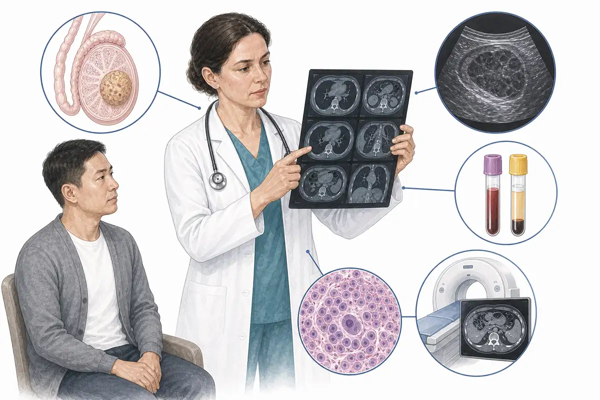
Basis for diagnosing testicular cancer
1.Scrotal ultrasound
Scrotal ultrasound is the preferred imaging modality for diagnosing testicular cancer; it can reveal hypoechoic solid masses within the testis and allow for the assessment of tumor size, margins, and blood flow signals.
2.Serum AFP test
Alpha-fetoprotein (AFP) levels are elevated in non-seminomatous germ cell tumors (particularly yolk sac tumors) but not in seminomas, making it a crucial marker for diagnosis and differential diagnosis.
3.Serum β-hCG test
Human chorionic gonadotropin (β-hCG) levels may be elevated in choriocarcinoma and seminoma; combined testing with alpha-fetoprotein (AFP) can assist in determining the tumor type.
4.Orchiectomy
High inguinal orchiectomy is the preferred method for diagnosing and treating testicular cancer, allowing for the retrieval of an intact specimen for pathological examination and staging.
5.LDH testing
Lactate dehydrogenase (LDH) levels are frequently elevated in testicular cancer; as a marker of tumor burden, LDH is incorporated into the international staging system (S1–S3).
6.Abdominal CT scan
Contrast-enhanced CT to evaluate retroperitoneal lymph node metastasis; lymphatic spread to retroperitoneal lymph nodes is the most common route of metastasis for testicular cancer.
7.Chest CT scan
Assesses for metastases in the lungs and mediastinal lymph nodes; a standard examination for initial staging.
8.Whole-body PET-CT
Assesses systemic metastasis; it offers advantages in detecting metastatic lesions of seminoma and can be used to evaluate treatment efficacy and monitor for recurrence.
9.Postoperative marker monitoring
After orchiectomy, AFP and β-hCG levels are monitored weekly until they return to normal; an abnormally prolonged half-life suggests the presence of metastases and serves as a key indicator for follow-up monitoring.
Daily Self-Check
Perform a testicular self-examination monthly by palpating the testicles to check for lumps or changes in firmness.Seek immediate medical attention if painless testicular enlargement or a lump is detected.
Conclusion
Diagnosis of testicular cancer relies on scrotal ultrasound and tumor marker testing. Cure rates are extremely high with early detection and treatment;men are advised to perform monthly testicular self-examinations.


