


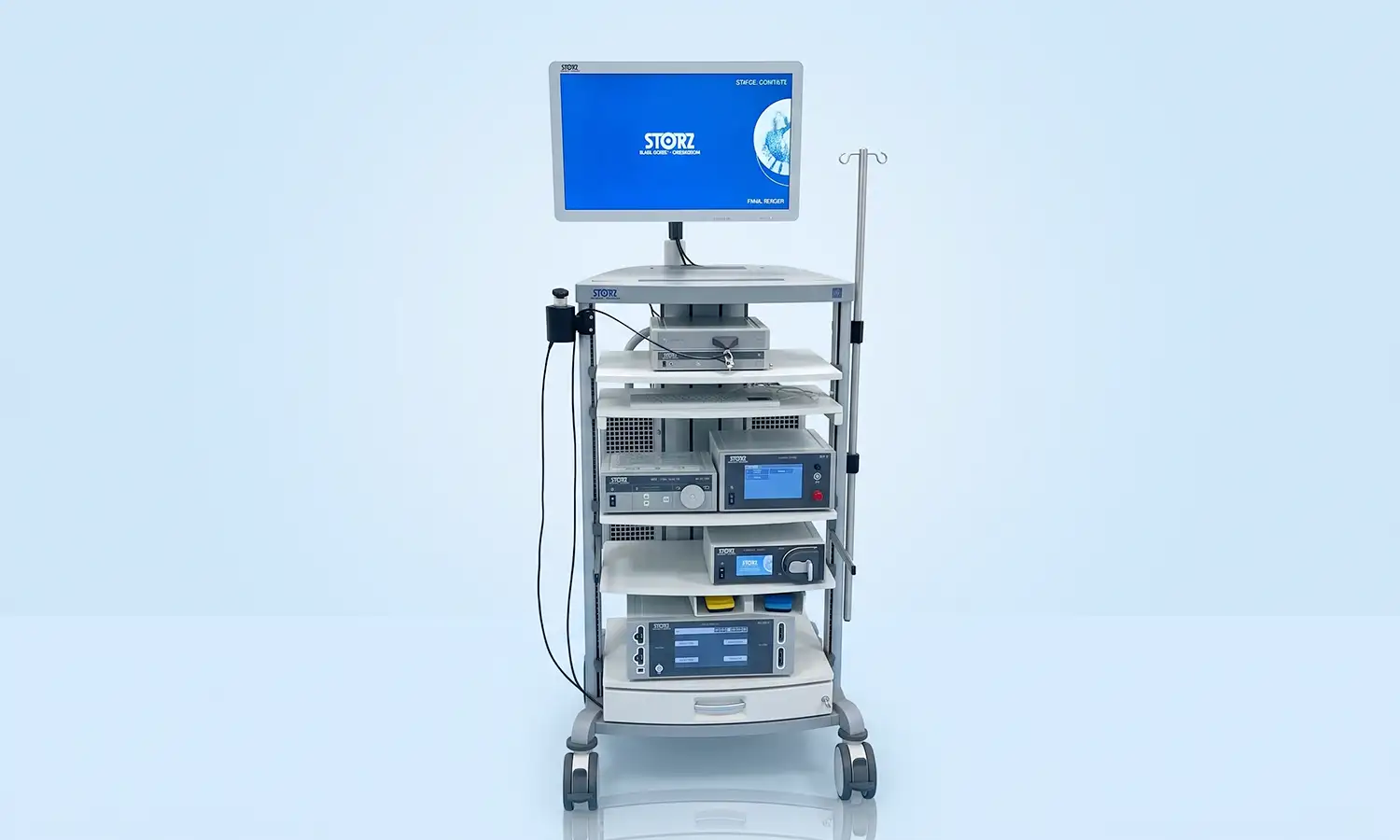
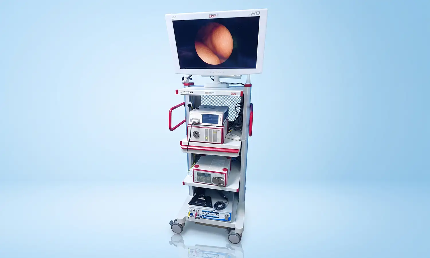
Laparoscopic systems are developed by internationally renowned endoscopy brands such as KARL STORZ, R. WOLF, and OLYMPUS, serving as the core imaging equipment for minimally invasive surgery. The system comprises components such as a high-definition camera system, a cold light source, an insufflator, a monitor, and various surgical instruments.By inserting a miniature camera and light source into the patient's body, the system transmits real-time images of internal organs to a display screen, enabling surgeons to perform precise surgical procedures while viewing the screen.
Core Technologies
What is a laparoscopic system?
The laparoscopic system is the core equipment for minimally invasive surgery.During the procedure, the surgeon makes two to four small incisions (0.5–1.5 cm) in the patient's abdomen and inserts a laparoscope equipped with a camera. A high-definition camera transmits real-time images of internal organs to a display screen.Surgeons perform precise surgical tasks—such as exploration, dissection, cutting, and suturing—while viewing the screen.Compared to traditional open surgery, laparoscopic surgery avoids large abdominal incisions, resulting in less trauma, reduced blood loss, and faster recovery for the patient.
1. High-definition and 3D stereoscopic imaging technology: Enabling doctors to "see more clearly"
Mainstream laparoscopic systems focus on three imaging modalities: 3D (three-dimensional), 4K ultra-high definition (UHD), and fluorescence.3D laparoscopes mimic human vision by using dual built-in cameras to capture simultaneous images; these are displayed using different polarizations to create binocular disparity, thereby generating a stereoscopic effect.When wearing 3D glasses, surgeons view a stereoscopic image of the body, gaining a realistic sense of depth and up to 4x magnification.Brands such as STORZ have introduced "three-in-one" laparoscopic systems that integrate 4K UHD, 3D stereoscopic vision, and fluorescence navigation, combining ultra-high image quality, stereoscopic depth perception, and real-time tissue visualization.
3D high-definition technology reconstructs internal 3D structures through minimal incisions, enabling maximum surgical precision, minimizing damage to blood vessels and nerves, and shortening operative time.This technology is particularly well-suited for delicate anatomical procedures, offering unique advantages when addressing complex areas such as high-level para-aortic lymph nodes and the ureteral tunnel.
2. Fluorescence navigation and precise positioning technology: Assists doctors in accurately locating the lesion.
Fluorescence laparoscopy utilizes fluorescent dyes, such as indocyanine green (ICG), to make lymphatic vessels, lymph nodes, and tumor-feeding blood vessels "glow" and become visible under the laparoscopic view.Surgeons can precisely identify the extent of the lesion to be resected, sentinel lymph nodes, and the course of critical blood vessels, achieving surgical outcomes characterized by "clearer visualization and greater resection precision."Intraoperative ultrasound and fluorescence imaging guide surgeons in performing precise procedures using advanced laparoscopic instruments.
3. Flexible instruments and minimally invasive techniques: Supporting complex surgical procedures
The laparoscopic system is equipped with a variety of precision surgical instruments, including ultrasonic scalpels and electrosurgical workstations.The hospital routinely performs reduced-port laparoscopic surgery, which further minimizes the number of incisions compared to traditional laparoscopy, achieving a more minimally invasive outcome.
Scope of Indications
High-definition/3D laparoscopic systems are widely used across various surgical fields, particularly for complex conditions requiring precise resection and functional preservation.
1. Clinical applications
• Hepatobiliary and pancreatic surgery
Primary Applications:
Laparoscopic liver cancer resection;
Pancreaticoduodenectomy;
Surgery for biliary tract diseases;
Treatment of benign and malignant hepatobiliary tumors.
Advantages:
Uses high-definition magnification and stereoscopic visualization to assist physicians in precisely managing complex anatomical structures, such as hepatic blood vessels and bile ducts.
• Gastrointestinal surgery
Primary Applications:
Radical gastrectomy for gastric cancer
Radical surgery for colorectal cancer;
Resection of gastrointestinal stromal tumors (GIST)
Advantages:
Reduces surgical trauma while meeting the requirements for complete tumor resection and lymph node dissection.
• Urology
Primary Applications:
Renal tumor resection;
Radical prostatectomy;
Bladder cancer surgery.
Advantages:
The high-definition 3D view helps surgeons precisely identify nerve and vascular structures, allowing for tumor treatment while preserving organ function.
• Gynecologic tumors
Primary Applications:
Radical surgery for cervical cancer;
Staging surgery for endometrial cancer;
Ovarian tumor resection.
Advantages:
Minimally invasive techniques reduce postoperative pain and accelerate recovery.
2. Suitable patient population
• Individuals requiring minimally invasive surgery for thoracic or abdominal tumors
For patients with tumors requiring surgery—such as liver, gastric, colorectal, renal, or cervical cancers—laparoscopic surgery enables precise tumor resection through a minimally invasive approach.
• Individuals seeking surgical options with less trauma and faster recovery
Compared to traditional open surgery, laparoscopic surgery requires only a few small incisions, resulting in less postoperative pain, shorter hospital stays, and faster recovery for patients.
• Patients requiring surgery involving fine anatomical dissection and functional preservation
For procedures requiring meticulous technique—such as radical prostatectomy with preservation of sexual and urinary continence functions, or kidney-sparing surgery for renal tumors—the stereoscopic view and magnification capabilities of 3D high-definition laparoscopy significantly enhance surgical precision.
Core Strengths
Comparison Dimension Conventional Open Abdominal Surgery HD/3D Laparoscopic System
Incision size Large abdominal incision (10–20 cm) 2–4 small punctures (0.5–1.5 cm)
Surgical Field of View Direct naked-eye view; limited visibility 4K Ultra-HD + 3D stereoscopic view; 4x magnification
Depth perception Natural stereoscopic vision 3D imaging with a realistic sense of depth
Operational precision Manual operation; subject to physiological tremor High-definition magnification and 3D depth perception; precise identification of blood vessels and nerves
Degree of trauma Extensive muscle dissection Minimally invasive; minimal tissue damage
Intraoperative bleeding Significant blood loss Significant reduction in blood loss
Postoperative recovery Long hospital stay Shorter hospital stay, faster recovery
Surgery time Standard Surgery time reduced for some procedures
Traditional open surgery
Traditional open surgery involves making a large incision to directly expose the surgical site, with the surgeon relying on direct visualization and manual manipulation to perform the procedure.While this offers a direct view, the large incisions required can lead to greater tissue damage, increased postoperative pain, a higher risk of bleeding, and longer recovery times.When operating in complex anatomical regions, the surgeon's ability to identify delicate structures is limited by the constraints of natural human vision and the available operating space.
High-Definition/3D Laparoscopic System
The high-definition/3D laparoscopic system accesses the surgical site through small, minimally invasive incisions. It replaces traditional direct visualization with 4K ultra-high-definition imagery and a 3D stereoscopic view, offering approximately 4x magnification and a realistic sense of depth. This enables surgeons to clearly identify blood vessels, nerves, and tissue planes.Compared to traditional open surgery, the laparoscopic system requires only a few small incisions, reducing tissue trauma and intraoperative bleeding while shortening postoperative recovery time.The combination of 3D stereoscopic imaging and high-definition magnification enhances surgical precision, allowing for finer execution of steps such as dissection, resection, and suturing.Additionally, certain minimally invasive procedures can reduce operative time and improve overall treatment efficiency.
Clinical Application Case
1. Colorectal tumors
Examinations revealed neoplastic lesions in the patient's ascending colon and sigmoid colon, requiring surgical resection.Traditional surgery typically involves an abdominal incision to extract the specimen, resulting in significant trauma and a prolonged recovery period.To minimize surgical trauma, the treatment team performed a fully laparoscopic colorectal tumor resection combined with the NOSES (Natural Orifice Specimen Extraction Surgery) technique.
• Treatment regimen
Minimally invasive surgery assisted by a high-definition/3D laparoscopic system; procedures include total laparoscopic right hemicolectomy, sigmoid colon tumor resection, and Natural Orifice Specimen Extraction Surgery (NOSES).
• Outcomes and follow-up
The surgery was successfully completed, allowing for specimen extraction without an additional abdominal incision.The patient recovered well postoperatively; compared to traditional open surgery and conventional laparoscopic procedures, the NOSES technique minimizes abdominal wall trauma, reduces postoperative pain, and helps shorten recovery time.
FAQ
1. What is the difference between 3D laparoscopy and standard laparoscopy?
3D laparoscopy provides the surgeon with a stereoscopic view featuring depth perception, facilitating the observation of anatomical planes and the execution of precise maneuvers.It remains a surgical tool operated by the surgeon; therapeutic outcomes depend on the patient's condition, the surgical procedure, and the team's experience.
2. Does 3D laparoscopic surgery require general anesthesia?
Most laparoscopic surgeries are performed under general anesthesia, with the camera and instruments inserted through small incisions in the abdominal wall.The specific anesthesia method, number of incisions, and duration of surgery vary depending on the surgical site and complexity.
3. Which diseases may be treated using 3D laparoscopic surgery?
3D laparoscopy can assist in performing select minimally invasive surgeries across various specialties, including general surgery, urology, and gynecology.Suitability is determined by a comprehensive assessment of factors such as the nature of the disease, the extent of the lesion, prior surgical history, physical condition, and the presence of any contraindications for minimally invasive surgery.
4. Does using 3D laparoscopy guarantee that conversion to open surgery won't be necessary?
No guarantees can be made.If severe adhesions, hemorrhage, unclear anatomy, or other safety-compromising conditions are encountered during the procedure, the physician may need to extend the incision or convert to open surgery; such decisions are made to ensure patient safety.
5. How can I inquire about 3D laparoscopic surgical treatment?
If you wish to determine your suitability for 3D laparoscopic surgery, please bring your diagnosis certificate, recent imaging, endoscopic or pathology results, past surgical records, and a list of current medications.The surgeon will evaluate surgical indications, anesthesia risks, and available surgical options, and explain the expected benefits and potential risks.