


Indications
1. Applicable conditions
• Early-stage solid tumors (regarding lesion size and number).• Patients with limited options—such as the elderly or those with poor cardiopulmonary or liver function—who cannot tolerate open surgical resection
• Patients with local recurrence (solitary or oligometastatic lesions) after surgery or radiotherapy/chemotherapy who require repeat local intervention.
• Systemic tumors presenting with localized metastatic lesions (e.g., oligometastases in the liver, lung, or kidney) where the primary tumor is under stable control.
• Intermediate-to-advanced stage tumors; used to alleviate symptoms and shrink lesions, in combination with interventional, targeted, and immunotherapies as part of a comprehensive treatment plan.
2. Common indications
Localized solid lesions such as primary liver cancer, liver metastases, peripheral lung cancer, lung metastases, renal cancer, adrenal tumors, bone metastases, thyroid microcarcinoma, and small breast nodules.
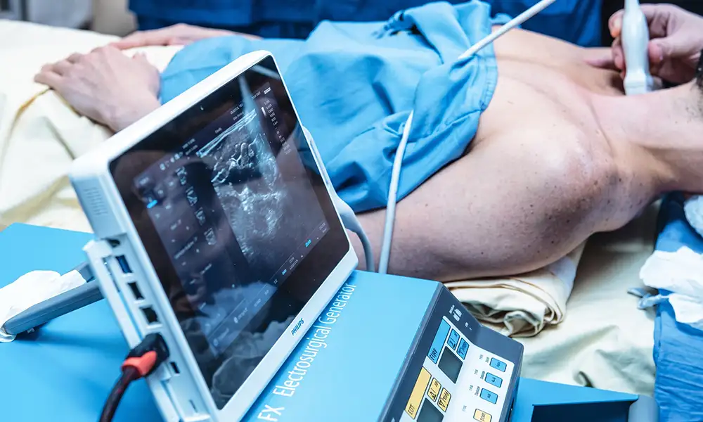
Core Strengths
1. Minimally invasive puncture; reduced physical burden
Requires only a millimeter-scale skin puncture tract;no thoracotomy or laparotomy is needed; local sedation is typically used during the procedure; the postoperative hospital stay is short;and it is better tolerated by elderly patients and those with multiple underlying conditions.
2. Real-time, image-guided precision temperature control for inactivation
CT and ultrasound provide continuous, synchronized monitoring of electrode placement, concentrating heat on the lesion while maintaining a safety margin to minimize the risk of thermal damage to surrounding healthy organs.
3. Suitable for special patient populations unable to undergo surgery
Provides alternative local intervention options for patients with liver cirrhosis, severe cardiopulmonary impairment, or physical frailty following multiple surgeries who are unable to undergo major operations.
4. Can be performed repeatedly
If new or residual lesions appear subsequently, repeat ablation sessions can be performed—provided organ function permits—to continuously manage the local tumor burden.
5. Highly adaptable comprehensive treatment plan
Can be combined with preoperative TACE to shrink the tumor and paired with postoperative targeted therapy, immunotherapy, and radiotherapy to establish an integrated systemic and local treatment plan.
Technical features
1. MDT: Upfront design of personalized treatment plans
Joint consultations involving interventional radiologists, oncologists, radiologists, and surgeons are held for every patient. Suitability for ablation is assessed based on lesion size, location, and organ reserve, followed by the planning of puncture paths and ablation margins.
2. Multimodal imaging navigation platform
Equipped with enhanced CT and ultrasound fusion localization systems to enable precise, layered planning for high-risk lesions located near major blood vessels, bile ducts, and the trachea, thereby avoiding the risk of thermal injury.
3. Standardized quality control system covering the entire process
Strict adherence to domestic expert consensus on thermal ablation: comprehensive pre-operative screening of coagulation and organ function; real-time intra-operative monitoring of temperature and puncture depth;and immediate post-operative imaging assessment of the ablation coverage area.
4. Integration of comprehensive diagnosis and treatment
The ablation center coordinates with interventional wards, medical oncology, and radiation oncology departments to schedule ablation and systemic therapies, thereby reducing the need for patients to travel between multiple departments.
5. Stratified, long-term follow-up mechanism
Scheduled contrast-enhanced imaging follow-ups at 1, 3, and 6 months post-procedure to dynamically monitor lesion changes and timely determine the need for supplementary ablation or adjustments to the systemic treatment plan.
Technical definition
Radiofrequency ablation (RFA) is a minimally invasive, image-guided thermal ablation technique. It involves inserting an electrode needle into the tumor under CT or ultrasound monitoring and using high-frequency alternating current to generate heat through the friction of tissue ions. This steadily raises the local temperature above 60°C, inducing coagulative necrosis in tumor cells. RFA is a key local intervention in the multidisciplinary comprehensive treatment of solid tumors, with procedures performed in accordance with national expert consensus guidelines on tumor thermal ablation.
Working principle/mechanism
High-frequency radiofrequency current is delivered to the lesion via electrodes, causing ions within the tissue to vibrate and rub against each other, generating continuous heat. When the local temperature is maintained at 60°C or higher, proteins within tumor cells denature and cell membranes rupture, resulting in irreversible damage. Simultaneously, the high heat seals off the tiny blood vessels supplying the lesion, cutting off the tumor's nutrient supply; this dual action inhibits the progression of the lesion. Real-time imaging monitoring is used throughout the procedure to precisely control the thermal treatment zone, preventing extensive burns to healthy tissue.
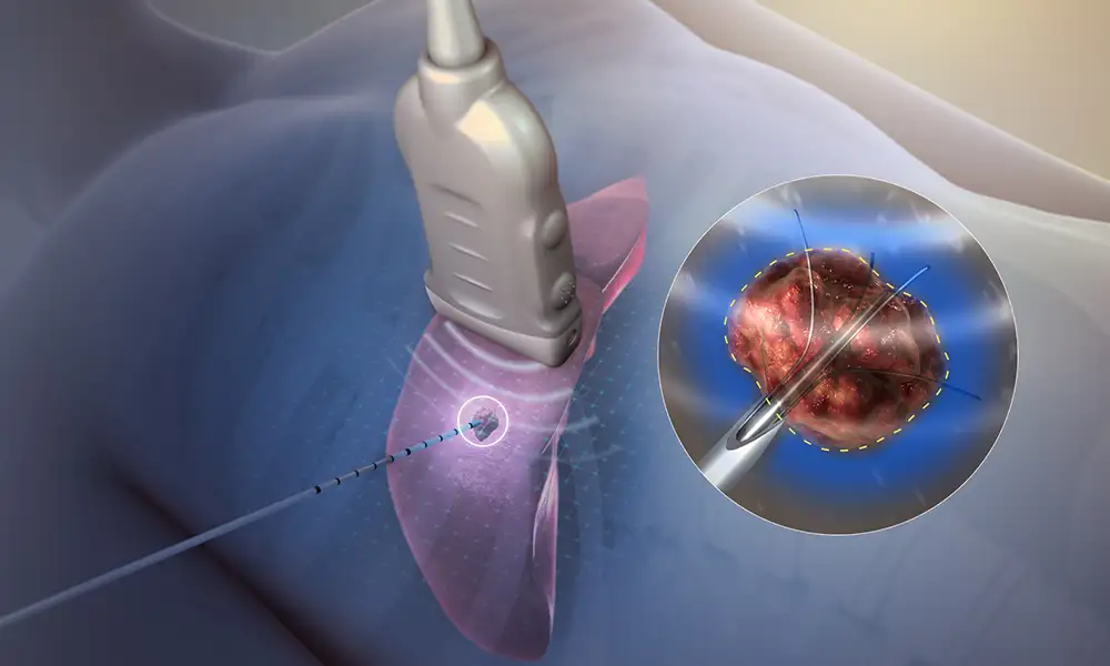
Treatment process
1. Comprehensive preoperative assessment: Complete contrast-enhanced CT/MRI, liver and kidney function tests, coagulation studies, and cardiopulmonary function tests; conduct an MDT consultation to confirm the feasibility of ablation and the puncture plan.
2. Image-guided localization and planning: CT/ultrasound scans mark the skin entry point and calculate a safe needle path, avoiding critical structures such as blood vessels, bile ducts, and the trachea.
3. Electrode placement via puncture under local anesthesia: Percutaneous puncture is performed under sedation and analgesia to precisely guide the electrode to the tumor target area.
4. Segmented radiofrequency (RF) thermal ablation: RF energy is delivered at multiple points and time intervals with real-time intraoperative temperature monitoring to ensure ablation margins meet standards.
5. Needle tract ablation for hemostasis and device withdrawal: Upon completion of ablation, the needle is withdrawn while simultaneously cauterizing the puncture tract to reduce the risk of bleeding and seeding; the puncture site is then pressure-bandaged.
6. Post-operative monitoring and scheduled follow-up: Hospital observation of vital signs and the puncture site for bleeding; periodic imaging re-examination to assess lesion inactivation status.
Safety and Precautions
1. MDT: Preoperative screening as the foundation for safety
Strictly screen out patients unsuitable for ablation—such as those with severe coagulation disorders, diffuse multiple lesions, severe organ failure, or active infections—and proactively assess the risks of puncture and thermal injury.
2. Continuous dynamic imaging monitoring throughout the procedure
Real-time monitoring of electrode position and temperature parameters; for lesions adjacent to sensitive organs, the duration of individual ablation sessions is reduced, and treatment is administered in multiple fractions and zones.
3. Transient normal post-operative reactions
Some patients may experience low-grade fever, dull pain at the puncture site, and mild fatigue; these are generally short-term stress reactions following ablation that gradually subside with symptomatic treatment.
4. Specialized care for the puncture site
Keep the post-procedure puncture site dry and clean; avoid rubbing or exposing the area to water in the short term. Refrain from lifting heavy objects or engaging in strenuous running or jumping for 1–2 weeks to prevent bleeding or hematoma at the puncture site.
5. Prompt follow-up visit for abnormal symptoms
If symptoms such as persistent high fever, severe chest or abdominal pain, massive hemoptysis or hematemesis (vomiting blood), or difficulty breathing occur, contact medical staff immediately for medical attention.
6. Unified coordination of comprehensive treatment
The timing intervals and medication cycles for combining ablation with interventional, targeted, or immunotherapies are determined by a multidisciplinary team; follow-ups and treatments must not be interrupted without medical advice.
FAQs
1. What is radiofrequency ablation?
Radiofrequency ablation involves placing an electrode needle into the lesion under imaging guidance and using high-frequency alternating current to raise the temperature of surrounding tissue, inducing coagulative necrosis to achieve local therapeutic goals.
2. Which lesions might be suitable for radiofrequency ablation?
Suitable for certain small, limited-number solid lesions in the liver, lung, kidney, or bone, as well as some benign nodules (e.g., thyroid).Selection is influenced by lesion size, location, and distance from blood vessels or nerves.
3. What are the differences between radiofrequency ablation, microwave ablation, and cryoablation?
Both RF and microwave ablation are forms of thermal ablation, though they differ in how energy is generated and propagated; cryoablation, conversely, utilizes freezing and thawing.When treating lesions near major blood vessels, blood flow may dissipate some of the heat; the choice of method depends on the specific characteristics of the lesion.
4. Does the treatment require general anesthesia?
Procedures may be performed under local anesthesia combined with sedation, or under general anesthesia for complex sites or special circumstances.Hospital observation is typically required following treatment of sites such as the lungs or liver;the duration depends on the risk of complications and the patient's recovery status.
5. Can ablation be performed again if the lesion was not completely covered in the first session?
If follow-up indicates local residual disease or new localized lesions, repeat ablation or combination therapy may be considered—provided organ function and conditions for needle puncture allow—though safety must be re-evaluated.
6. How is the treatment efficacy assessed?
Assessment cannot be based solely on pain relief or short-term tumor shrinkage.It typically requires evaluation via contrast-enhanced CT, MRI, or ultrasound to determine whether the ablation zone completely covers the lesion and whether any abnormal enhancement persists.


