


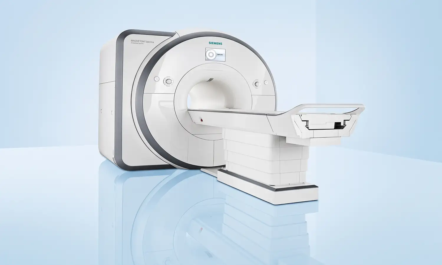
Developed by Siemens Healthineers (Germany), the MAGNETOM Vida 3.0T MRI system represents a new generation of premium 3.0T MRI technology. It features the industry's first BioMatrix Technology—integrating the latest in biotechnology, smart sensors, and AI—marking a milestone in MRI development over the past two decades.Unlike traditional MRI systems that require strict patient cooperation, the Vida system senses human anatomy, physiological data, and magnetic field homogeneity the moment the patient lies on the scanning table, enabling an intelligent examination process where the "equipment adapts to the patient."
Core Technologies
What is the Vida 3.0T MRI scanner?
Magnetic Resonance Imaging (MRI) is an imaging technique characterized by multi-planar and multi-parameter capabilities, a wide field of view, and the absence of ionizing radiation. It offers high image contrast and spatial resolution, providing excellent morphological and rich functional information for disease diagnosis.MAGNETOM Vida, launched by Siemens Healthineers, is the world's first 3.0T MRI system featuring "BioMatrix" technology (vital sign sensing).By integrating biotechnology, smart sensors, and artificial intelligence, it senses individual patient data—such as respiration, heartbeat, and anatomical structure—in real-time during the scan and automatically adjusts scanning parameters. This allows for the acquisition of high-quality diagnostic images without requiring the patient to make a conscious effort to cooperate.
1. BioMatrix Technology (Vitality Sensing System)
The Vida is equipped with the industry's first BioMatrix system, which senses physiological information—such as patient anatomy, respiratory rhythm, heart rate, and magnetic field variations—in real-time and relays this data to the entire MRI system.This "system-adapts-to-patient" scanning philosophy breaks the traditional constraint where the patient had to adapt to the equipment; patients can undergo body scans while breathing freely, without the need for respiratory gating devices or forced breath-holding.For cardiac examinations, the BioMatrix system enables the industry's fastest cardiac cine imaging, performing 10 to 20 times faster than conventional methods.
2. Intelligent, ultra-fast imaging and patient-comfort design
The Vida incorporates Turbo Suite acceleration technology and Deep Resolve deep-learning image reconstruction, boosting scan speeds by up to 91% compared to conventional systems.It features an industry-leading 70cm wide-bore design and the Quiet Suite platform, which reduces scan noise by 70% to 99%.The Tim 4G ultra-high-density coil platform offers up to 204 receive channels, making it one of the systems with the highest channel counts in the industry.Furthermore, EcoPower intelligent power management reduces energy consumption by 30% during standby.
Scope of Indications
The clinical applications of the Vida 3.0T MRI cover all body systems. In addition to conventional morphological imaging, it supports various functional imaging techniques, such as DWI, MRS, DTI, SWI, BOLD, and perfusion imaging.
1. Major clinical applications
Field Primary Applications
Tumor diagnosis and treatment; whole-body PET-based tumor screening, precision staging, treatment efficacy assessment, recurrence monitoring, and detection of micro-lesions
Central Nervous SystemEarly diagnosis of brain tumors, stroke, and neurodegenerative diseases (such as Alzheimer's disease)
Cardiovascular system Assessment of cardiac structure, function, and myocardial viability without the need for contrast agents
Musculoskeletal system: Imaging of fine structures (articular cartilage, ligaments, tendons, etc.), diagnosis of sports injuries
Abdominopelvic System Precision diagnosis of liver cancer, pancreatic cancer, prostate cancer, uterine and ovarian tumors, etc.
2. Suitable patient population
• Individuals requiring early tumor screening or regular follow-up examinations
The Vida supports techniques such as whole-body PET-like imaging and diffusion-weighted imaging (DWI), allowing for the assessment of whole-body metastases in a single scan.As MRI involves no ionizing radiation and poses no harm to the body, it serves as an ideal follow-up tool for oncology patients requiring repeated examinations.
• Individuals unable to cooperate with conventional MRI scans
Children, the elderly, and patients with breathing difficulties or claustrophobia often struggle to comply with the requirements of traditional MRI scans, such as lying still for long periods and repeatedly holding their breath.The Vida system's 70cm wide bore and ultra-quiet design significantly alleviate claustrophobia, while free-breathing scanning technology allows frail patients to complete the examination without the need for deliberate cooperation.
• Patients with complex cases requiring multimodal functional imaging
For patients requiring assessment of tissue metabolism, perfusion, and diffusion, the Vida system performs a comprehensive range of functional imaging in a single session, providing extensive data for precise diagnosis.
• Patients with cardiovascular or neurological diseases
Vida’s non-invasive cardiac cine imaging and brain functional imaging technologies provide precise, non-invasive imaging data for diagnosing conditions such as coronary heart disease, cardiomyopathy, brain tumors, and stroke.
Core Strengths
Comparison Dimension Conventional MRI Vida 3.0T MRI
Technical philosophy Patient adapts to equipment Equipment adapts to the patient; real-time sensing of physiological data allows for automatic adjustments
Aperture: Approx. 60 cm; 70 cm extra-large aperture for significantly improved comfort
Noise Relatively high Ultra-quiet platform; reduces sound pressure by 70%–99%
Scan speed Relatively slow AI-driven ultra-fast imaging; speed increased by up to 91%
Respiratory cooperation Requires strict breath-holding Free-breathing scanning; no respiratory gating device required
Coil channels; fewer; 204-channel Tim 4G (highest number of receive channels in the industry)
Cardiac imaging Requires heart rate control and breath-holding Free-breathing cardiac cine imaging; speed increased by 10–20 times
Functional imaging Limited Multimodal functional imaging (DWI, MRS, DTI, SWI, ASL, etc.)
Conventional MRI Examination
Conventional MRI provides high-quality images of tissue structures and serves as a vital tool for clinical diagnosis.However, the procedure requires patients to maintain a fixed position for an extended period and may necessitate maneuvers such as breath-holding.Factors such as the scanner's bore size, noise levels, and scan duration can cause anxiety, discomfort, or difficulty in cooperation for some patients.Regarding imaging capabilities, conventional MRI focuses primarily on structural imaging and has limitations in the multidimensional functional assessment of complex diseases; furthermore, examinations of the heart and dynamic organs often place high demands on patient cooperation.
Vida 3.0T MRI
The Vida 3.0T MRI adopts a patient-centric, intelligent design philosophy; it enhances examination adaptability and stability by sensing the patient's physiological state in real-time and automatically adjusting scanning parameters.The device features an extra-large bore (approximately 70 cm)—compared to the traditional 60 cm bore—which improves patient comfort and reduces feelings of claustrophobia.Regarding efficiency, the Vida 3.0T integrates AI-assisted imaging technology to significantly increase scanning speeds, with imaging efficiency for certain sequences improving by up to approximately 91%.Additionally, ultra-quiet technology reduces noise during the examination, thereby improving the patient experience.For complex imaging, the system features 204-channel Tim 4G coil technology to enhance signal acquisition capabilities. It supports multimodal functional imaging techniques—such as DWI, MRS, DTI, SWI, and ASL—providing comprehensive imaging data for oncology, neurology, and cardiovascular applications.Additionally, the Vida 3.0T supports free-breathing dynamic cardiac imaging, eliminating the need for breath-holding and improving the stability of cardiac and dynamic organ examinations.
FAQ
1. Does a 3.0T MRI scan involve radiation?
MRI utilizes strong magnetic fields and radiofrequency signals for imaging; it does not use X-rays and therefore produces no ionizing radiation.However, strict safety requirements regarding magnetic fields still apply, and suitability for the procedure must be assessed based on any internal implants and the patient's specific condition.
2. Can I undergo an MRI if I have metal objects or implanted devices in my body?
It is necessary to first verify the exact name, model, and MRI compatibility specifications of the implant.Cases involving cardiac pacemakers, cochlear implants, aneurysm clips, or metallic foreign bodies cannot be assessed based on general experience alone; patients should provide proof of implantation or surgical records so that professionals can conduct an evaluation and make a determination.
3. What if I am afraid of enclosed spaces or cannot lie flat for extended periods?
Vida features a wider bore, which can improve the sense of space for some patients; however, the procedure still requires entering the magnet and remaining as still as possible.If you suffer from severe claustrophobia or pain, or are unable to lie flat, please inform the staff when scheduling your appointment so that options such as having a companion present, sedation, or alternative imaging protocols can be evaluated.
4. Is a contrast-enhanced MRI scan safe?
Contrast-enhanced MRI may involve the use of gadolinium-based contrast agents;while most patients tolerate them well, adverse reactions can still occur, and special assessment is required for patients with severe renal impairment.The decision to use contrast enhancement rests with the physician; patients should disclose any history of kidney disease, allergies, pregnancy, or breastfeeding prior to the examination.
5. How do I schedule a Vida 3.0T MRI scan?
Before booking an appointment, the attending physician usually needs to specify the area to be examined and the purpose of the scan.Please proactively disclose any internal implants, metal foreign bodies, pregnancy, claustrophobia, or renal function issues, and provide relevant documentation and previous imaging; staff will use this information to conduct a safety screening and advise on preparation requirements, such as fasting.