


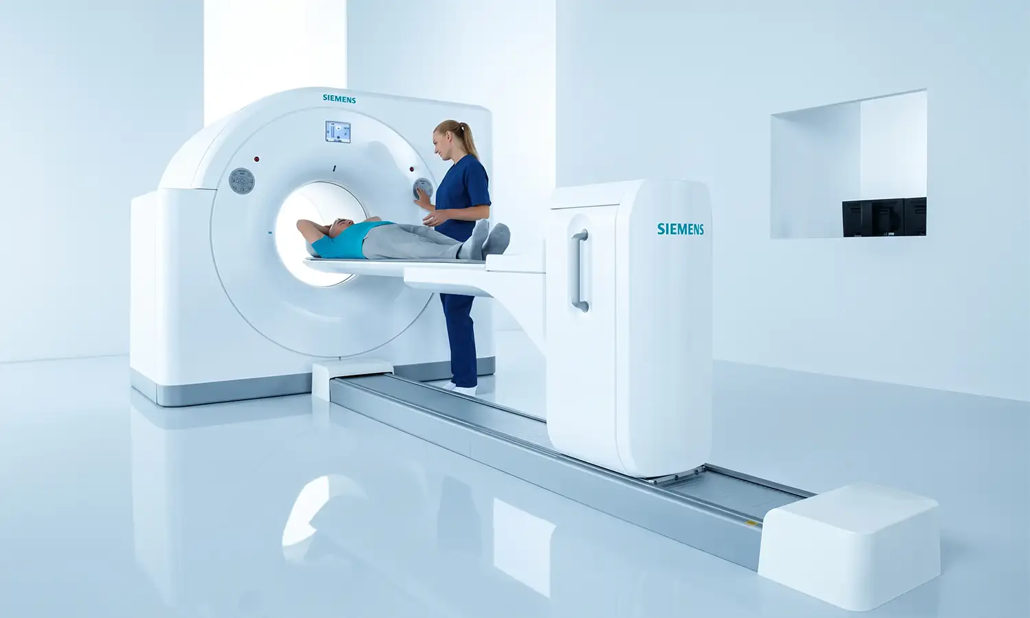
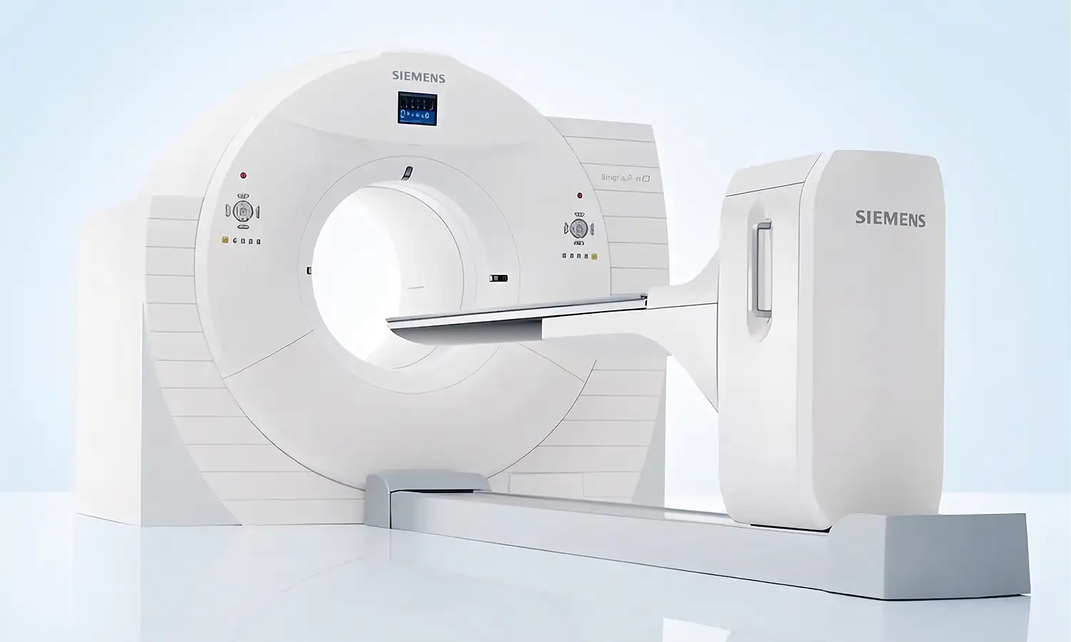
Developed by Siemens Healthineers (Germany), the Biograph mCT128 PET/CT is a high-end molecular imaging system that integrates Positron Emission Tomography (PET) with Computed Tomography (CT). The PET component reflects cellular metabolic activity, while the CT component precisely localizes anatomical structures;their combination enables dual diagnostic capabilities—characterizing the nature of a tumor ("qualitative" diagnosis) and pinpointing its exact location ("locational" diagnosis).
Core Technologies
What is PET/CT?
PET/CT is an integrated system that combines two imaging technologies: PET (Positron Emission Tomography) and CT (Computed Tomography).PET involves injecting a trace amount of a radiotracer (such as 18F-FDG) to visualize cellular metabolic activity; highly active tumor cells appear as high-uptake "hot spots" on the image. Meanwhile, CT provides precise anatomical localization.By fusing these images, physicians can simultaneously identify "where metabolic abnormalities exist" and "the specific anatomical location of those abnormalities," enabling precise tumor localization and characterization.
1. High-resolution LSO crystal detection technology
The Biograph mCT128 utilizes Siemens' LSO ultra-fast pure crystals, which are impurity-free, feature short decay times, and offer high photoelectric conversion efficiency, achieving an industry-leading resolution of 2 mm.This superior resolution ensures that even small, early-stage lesions can be clearly visualized.Equipped with mature technologies such as TrueV, HD·PET, and TOF (Time-of-Flight), the system can complete a standard whole-body PET/CT scan within 10 minutes; the CT component acquires up to 128 slices, enabling a whole-body CT scan in under 10 seconds.
2. Eco-friendly, low-dose technology
The Biograph mCT128 utilizes Siemens' proprietary "green" low-dose technology. Its X-ray tube employs direct oil cooling for the anode target, featuring a massive 50 MHU heat capacity that eliminates the need to pause for cooling.Offering lower radiation doses and shorter scan times, it is truly a "green" system.It features the industry's only 78cm large-bore design, providing a more comfortable examination experience for larger patients or those with claustrophobia.
Scope of Indications
The clinical applications of the Biograph mCT128 PET/CT encompass the early detection and diagnosis of major diseases affecting the oncology, neurology, and cardiology fields.
1. Major clinical applications
Field Primary Applications
Tumor diagnosis and treatment; differential diagnosis of benign and malignant tumors, tumor staging, monitoring and assessment of treatment efficacy, recurrence detection, and identification of primary tumor sites
Neurological system: localization of epileptic foci, early diagnosis of Alzheimer's disease, etc.
Cardiovascular system Myocardial viability assessment, etc.
High-end health check-up | Early-stage tumor screening
2. Suitable patient population
• Individuals requiring early tumor screening or regular follow-up examinations
A single PET/CT scan enables whole-body imaging, providing a sensitive assessment of systemic metabolic activity.In 2022, this equipment assisted in the precise screening and diagnosis of tumor lesions for over 2,100 patients.
• Cases where differentiating between benign and malignant tumors is difficult
PET/CT significantly improves diagnostic accuracy by enabling a "four-dimensional" diagnosis—determining the disease's location, nature (benign vs. malignant), quantitative characteristics, and stage.Diagnostic accuracy can exceed 90%.
• Patients requiring assessment of treatment efficacy
PET/CT enables precise assessment of the efficacy of radiotherapy, chemotherapy, targeted therapy, and immunotherapy; it allows for the timely evaluation of treatment effectiveness and provides an objective basis for adjusting treatment plans.
• Patients with metastatic tumors of unknown primary origin
For cancer patients with identified metastases but an unknown primary tumor, whole-body PET/CT imaging can effectively locate the primary lesion.
Core Strengths
Comparison Dimension Conventional CT/MRI Biograph mCT128 PET/CT
Diagnostic Information: Simple anatomical structures vs. dual metabolic and anatomical information (fusion of function and structure).
Whole-body examination Requires multiple scans of different body regions Whole-body imaging completed in a single scan
Scan time Relatively long Whole-body PET/CT takes approximately 10 minutes; the CT component takes only 10 seconds
Resolution — 2mm; early-stage, minute lesions are clearly distinguishable
Radiation dose Relatively high Eco-friendly low-dose technology; lower radiation
Examination comfort Small aperture 78 cm large aperture; high level of comfort
Conventional CT/MRI Examinations
Conventional CT and MRI examinations primarily visualize tissue structures to reveal the location, size, and morphology of tumors. However, as they mainly reflect anatomical information, their ability to detect certain early-stage lesions or functional changes is limited.Whole-body screening often requires multiple sessions based on the body regions being examined, resulting in a relatively long process.Additionally, the small bore size of some conventional equipment can compromise patient comfort, particularly for larger patients or those prone to claustrophobia.
Biograph mCT128 PET/CT
Compared to traditional imaging methods, the Biograph mCT128 PET/CT enables whole-body imaging in a single scan; a full PET/CT exam takes approximately 10 minutes, with the CT component taking as little as 10 seconds.The system features a spatial resolution of approximately 2 mm, aiding in the detection of tiny, early-stage lesions.It employs low-dose imaging technology to reduce radiation exposure while maintaining image quality.Additionally, the 78 cm large-bore design enhances spatial comfort during the procedure, providing a more convenient and comfortable experience for patients.
FAQ
1. What is PET/CT primarily used to examine?
By integrating metabolic or molecular functional data with CT anatomical imaging, PET/CT is used for staging, assessing treatment efficacy, and monitoring recurrence in certain tumors, as well as for evaluating specific neurological and cardiovascular diseases.The decision to undergo the examination should be made by a physician based on specific clinical issues.
2. Why is fasting usually required before a PET/CT scan?
The distribution of commonly used tracers can be influenced by blood glucose levels, food intake, and physical activity; therefore, patients are typically required to fast, avoid strenuous exercise, and remain calm prior to the examination, in accordance with appointment instructions.Patients with diabetes should not discontinue their medication on their own;instead, they should inform the staff in advance so that medical personnel can coordinate the medication schedule with the examination time.
3. Does a PET/CT scan involve radiation?
PET/CT involves the use of a small amount of radioactive tracer and a CT scan, resulting in some exposure to ionizing radiation. Physicians select the appropriate protocol based on the clinical objective;patients who are pregnant, potentially pregnant, or breastfeeding should proactively inform the medical staff prior to scheduling the examination.
4. Why is it necessary to wait quietly after the tracer injection?
After the tracer is injected, it requires time to distribute throughout the body. During this waiting period, patients should generally rest quietly, minimize talking, and avoid walking around to reduce uptake by non-target tissues (such as muscles).Please follow the specific instructions provided by the imaging center regarding waiting times and fluid intake.
5. How do I schedule a Biograph mCT128 PET/CT scan?
Before making an appointment, the attending physician usually needs to clarify the purpose of the examination and issue the corresponding requisition.Please prepare past pathology reports, recent imaging (such as CT or MRI scans), previous PET/CT results, and treatment records. Additionally, inform staff in advance regarding your blood glucose levels, pregnancy or breastfeeding status, and recent medications so they can schedule the examination and explain necessary preparations.