


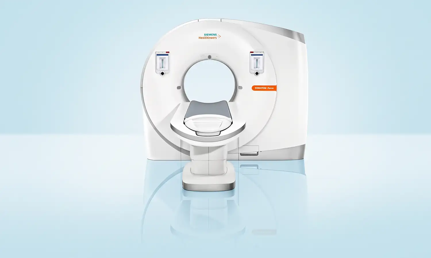
Developed by Siemens Healthineers (Germany), Dual-Source CT is currently one of the most advanced CT imaging systems in the world.Unlike traditional CT scanners equipped with only a single X-ray tube and detector, Dual-Source CT highly integrates two X-ray tube and detector systems within a single gantry.These two systems operate synchronously—effectively doubling the scanning speed—thereby achieving comprehensive improvements in scanning speed, image quality, and safety performance.
Core Technologies
What is a dual-source CT scanner?
Dual-source CT (DSCT) is a CT system that acquires images of the human body simultaneously using two independent X-ray tube and detector sets.While conventional CT utilizes a single X-ray tube and detector array, dual-source CT is equipped with two identical X-ray generation and detection systems mounted at an angle within the same gantry.These two systems can emit X-rays at either identical or different voltages, allowing for the integration or separation of data.
1. Ultra-fast dual-source scanning
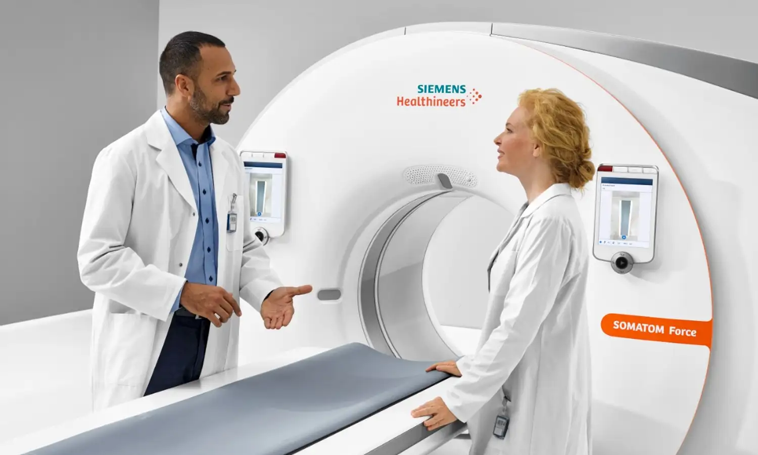
The dual-source CT system features two independent X-ray tube and detector sets working synchronously, doubling the scanning speed and imaging efficiency compared to conventional CT.It boasts the industry's fastest scanning speeds: a whole-heart scan can be completed in 0.25 seconds, whole-body major vessel imaging in 1 second, and a full aortic CTA examination in approximately 4 seconds.Scan speeds range from 458 mm to 737 mm per second—several times faster than conventional high-end CT scanners.This speed overcomes limitations imposed by physiological motion such as blood flow, respiration, and heartbeat, allowing elderly patients to undergo examinations without breath-holding and infants without sedation.Additionally, it incorporates spectral shaping technology (tin filtration) to filter out ineffective photons from the X-ray spectrum; the radiation dose for a single chest scan can be reduced to the sub-millisievert level—comparable to a standard chest X-ray and approximately 90% lower than conventional CT—while contrast agent usage is only one-quarter of that required for conventional CT.This technology enhances safety for early tumor screening and regular follow-up examinations.
2. Dual-energy material differentiation.
The two X-ray tubes in a Dual-Source CT system can be configured to scan simultaneously at different energy levels (e.g., low kV and high kV), acquiring two sets of data at different energies.The system enables quantitative separation and imaging of various materials—such as bone, water, fat, and iodine—to identify distinct tissue compositions.In tumor examinations, this unique dual-energy imaging capability enhances lesion detection rates and the ability to differentiate between benign and malignant tumors.
Scope of Indications
Dual-Source CT has clinical applications across all body systems, offering significant advantages particularly in early tumor screening, precise diagnosis, and the assessment of treatment efficacy.
1. Areas for examination/diagnosis
Field Primary Applications
Tumor diagnosis and treatment; whole-body early tumor screening, differentiation between benign and malignant tumors, precise staging, treatment efficacy assessment, and follow-up monitoring
Cardiovascular system Coronary CTA (no heart rate control required); "triple rule-out" (chest pain) one-stop examination
Neurological system: head and neck vascular imaging, one-stop stroke assessment.
Thorax and Abdomen: Pulmonary nodule screening and follow-up, precise diagnosis of liver lesions
Bone and joints: Non-invasive diagnosis of gouty tophi, metal artifact reduction
2. Suitable patient population
• Individuals requiring early tumor screening or regular follow-up examinations
The ultra-low radiation dose of Dual-Source CT makes tumor screening and regular follow-ups safer.It achieves a sensitivity of up to 94% for pulmonary nodule detection, while the radiation dose for a single chest scan is equivalent to that of a standard chest X-ray.
• Individuals unable to cooperate with conventional CT scans
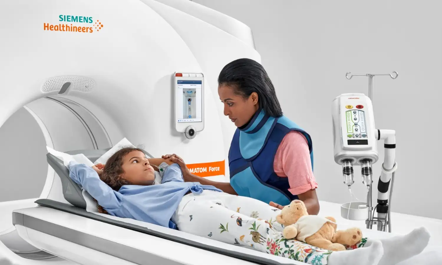
Elderly patients, children, and those with breathing difficulties can complete the examination without holding their breath.Infants and young children do not require sedation prior to the examination.
• Individuals requiring extensive or multi-site combined examinations
Combined head and neck examination takes less than 2 seconds; a whole-body scan takes as little as 4 seconds.One-stop imaging of multiple body regions can be completed in a single scan.
• Tumor patients requiring multiple follow-up imaging examinations
Cancer patients require multiple follow-up CT scans during treatment to evaluate therapeutic efficacy.Dual-source CT minimizes the radiation dose per scan, significantly reducing the cumulative radiation risk.
Core Strengths
Comparison Dimension Conventional CT Dual-source CT
X-ray tube/detector 1 set 2 sets, simultaneous acquisition
Scan speed Relatively slow Whole-heart scan completed in 0.25 seconds; whole-body major vessel imaging completed in 1 second
Temporal resolution Approx. 150 ms 66–75 ms
Radiation dose Relatively high Reduced by approx. 90% compared to conventional CT; equivalent to a single chest X-ray
Breath-holding requirement: Usually required vs. No breath-holding required (performed during free breathing)
Heart rate limitation Heart rate control required No heart rate control required; imaging possible even for patients with atrial fibrillation
Energy Imaging: Single-energy | Dual-energy (enables material separation and compositional analysis)
Tumor diagnosis; primarily morphological; utilizing dual morphological and functional data to improve differential diagnostic capabilities
Conventional CT Examinations
Conventional CT examinations acquire images using a single X-ray tube and detector array, clearly displaying anatomical structures. However, imaging rapidly moving structures—such as the heart and major blood vessels—demands high scanning speeds and temporal resolution.Some procedures require patients to hold their breath, and image quality can be compromised in patients with significant heart rate variability.Regarding tumor diagnosis, conventional CT focuses primarily on morphology, offering limited capability to analyze lesion function or composition.
Dual-Source CT
By utilizing synchronous data acquisition technology with dual X-ray tubes and dual detectors, Dual-Source CT enhances scanning speed and temporal resolution; it can complete a cardiac scan in approximately 0.25 seconds and perform rapid whole-body imaging of major blood vessels.With a temporal resolution of 66–75 milliseconds, it captures clearer images of rapidly moving organs and reduces the need for strict heart rate control or patient breath-holding.
Regarding radiation control, the dual-source CT system employs advanced low-dose technology to minimize radiation exposure while maintaining image quality.The equipment supports dual-energy imaging, enabling material separation and compositional analysis, which provides additional imaging data for the differential diagnosis of diseases.In tumor assessment, dual-source CT goes beyond monitoring morphological changes in lesions; by incorporating functional and compositional data, it enhances the capability to detect early-stage lesions and achieve precise diagnoses.
FAQ
1. What is the difference between dual-source CT and standard CT?
Equipped with two X-ray sources and detector systems, dual-source CT improves data acquisition speed and temporal resolution while enabling certain dual-energy imaging capabilities.It can be particularly beneficial for imaging the heart and major vessels, as well as for patients requiring rapid scanning;however, the decision to use specific modes rests with the physician based on the clinical purpose of the examination.
2. Is contrast agent injection required for a dual-source CT scan?
Not necessarily.Non-contrast scans typically do not use iodine-based contrast agents, whereas contrast-enhanced CT or angiography may require intravenous injection.The decision to use contrast depends on the clinical question being addressed, not merely on the type of equipment.
3. Is breath-holding or heart rate control required for a dual-source CT scan?
Thoracic, abdominal, or cardiac examinations often require brief breath-holding to minimize motion artifacts.For certain cardiac exams, the need for heart rate control is determined by the physician based on the imaging protocol, heart rhythm, and patient condition; please do not take heart-rate-lowering medication on your own.
4. Does dual-source CT involve higher radiation exposure?
CT scans involve ionizing radiation, but the actual dose depends on the area examined, the scan range, body habitus, and the imaging mode; the use of dual X-ray tubes does not necessarily imply a higher radiation dose. The medical team selects an appropriate dose control strategy based on diagnostic requirements.
5. How do I schedule a Siemens dual-source CT scan?
Before booking, the attending physician typically needs to issue a CT scan request based on clinical indications.Please prepare previous imaging and relevant test results, and specify whether contrast enhancement is required. Pregnant women (or those who might be pregnant) and individuals with a history of adverse reactions to iodine-based contrast agents, renal dysfunction, thyroid disease, or diabetes should inform staff in advance.