


Indications
1. Applicable conditions
• Lesions immediately adjacent to sensitive organs such as the spinal cord, optic nerve, gastrointestinal tract, or major blood vessels, requiring strict limitation of radiation dose to surrounding tissues
• Tumors in the chest or abdomen (e.g., lung, liver, pancreas) subject to respiratory motion, in patients unable to cooperate with treatment by holding their breath for extended periods.
• Solitary or multiple intracranial metastases and postoperative residual lesions requiring precise stereotactic fractionated irradiation
• Patients with solid tumors requiring systemic therapy for various objectives, including preoperative neoadjuvant treatment, postoperative adjuvant treatment, or palliative symptom relief
2. Common indications
• Head and neck region: nasopharyngeal carcinoma, primary/metastatic intracranial tumors, oromaxillofacial tumors
• Thorax: Lung cancer, breast cancer, esophageal cancer
• Abdominal/Digestive: Gastric cancer, rectal cancer, primary liver cancer
• Gynecologic tumors: cervical cancer, endometrial cancer
• GenitourinaryIndication: Prostate cancer
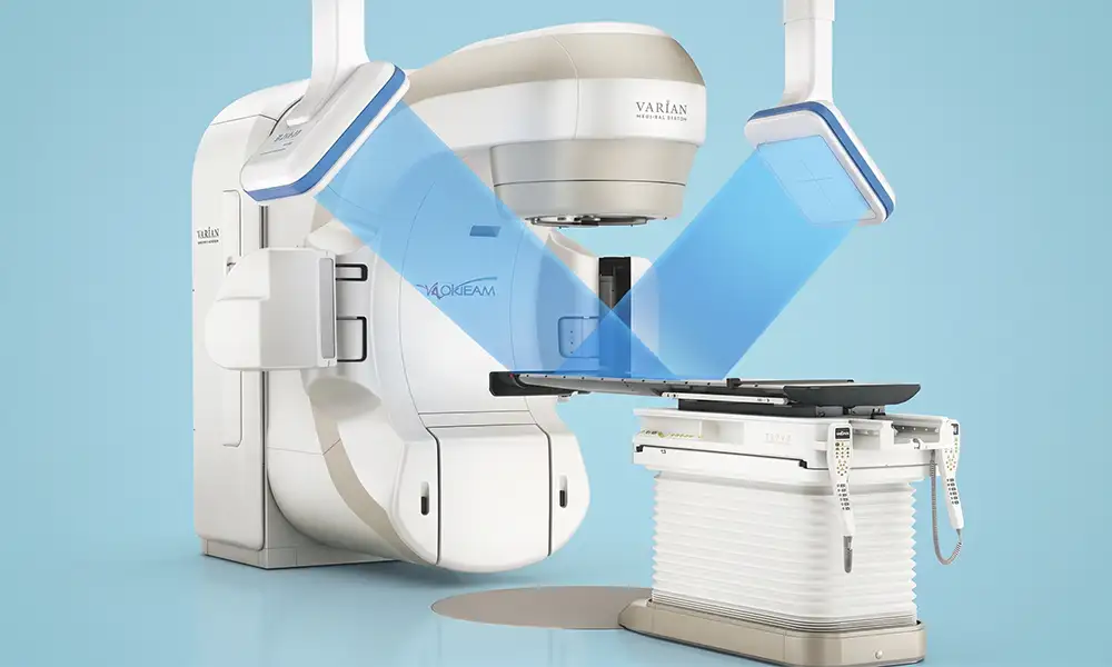
Core Strengths
1. 4D respiratory gating with free-breathing tracking
Features infrared surface sensing and 4D-CBCT imaging systems; patients do not need to hold their breath, as the system automatically identifies the stable respiratory phase for the tumor and delivers radiation only when the target area is correctly positioned. This significantly reduces radiation exposure to normal tissues—such as the heart and lungs—and improves tolerability for elderly or frail patients.
2. RapidArc high-speed rotational intensity modulation, reducing time spent on the treatment table
The gantry rotates 360° continuously with synchronized dynamic beam adjustment, compressing the single irradiation session to 3–5 minutes. This reduces soreness and discomfort caused by prolonged immobilization, making it suitable for patients with bone metastasis pain or limited mobility.
3. Real-time automatic correction via 6-degree-of-freedom treatment couch
Before each treatment session, Cone-Beam CT (CBCT) imaging is used to match the planning images, and a six-degree-of-freedom treatment couch automatically corrects for translational and rotational offsets. Precision self-checks are performed every 10 milliseconds throughout the treatment, keeping positioning errors within the sub-millimeter range.
4. One-stop compatibility with full-range radiotherapy techniques
Supports modalities including 3D-CRT (3D Conformal Radiotherapy), IMRT (Intensity-Modulated Radiotherapy), RapidArc (Volumetric Modulated Arc Therapy), IGRT (Image-Guided Radiotherapy), and SBRT (Stereotactic Body Radiotherapy), with physicians customizing dose plans based on lesion morphology and location.
5. Multi-level radiation settings to cover both superficial and deep lesions
Configured with multi-photon and multi-electron energy beams, suitable for treating both superficial skin tumors and deep-seated visceral tumors, covering a wider range of disease types.
Technical features
The radiotherapy center is equipped with the Vital Beam intelligent linear accelerator as a standard feature; the entire workflow is implemented in strict accordance with domestic expert consensus on precision radiotherapy:
1. MDT: Multidisciplinary upfront assessment
Joint consultations involving radiation oncologists, medical oncologists, radiologists, and surgeons are conducted for every patient to determine the timing of radiotherapy within surgical, targeted, and immunotherapeutic regimens. The overall treatment schedule is coordinated to ensure a comprehensive approach rather than relying solely on radiotherapy.
2. Closed-loop quality control system based on 4D imaging
Utilizes 4D-CT fused with MRI for target area localization; dose plans undergo dual-review by medical physicists; radiation limits for organs at risk—such as the lens, spinal cord, and gastrointestinal tract—are strictly enforced; and quality control checkpoints are established across the entire process of localization, planning, and treatment.
3. Patient distribution across multiple devices ensures treatment continuity
Staggered scheduling of multiple radiotherapy units minimizes the risk of treatment interruptions caused by equipment maintenance or patient surges, ensuring that fractionated radiotherapy is completed on schedule and maintaining a stable cumulative radiation dose.
4. Stratified, long-term follow-up management
Upon completion of the treatment course, a dedicated follow-up record is established. Periodic imaging re-examinations monitor changes in the lesion, while concurrent monitoring tracks radiation-induced reactions in the skin and digestive tract; symptomatic interventions and subsequent systemic treatment plans are adjusted as needed.
Technical definition
Vital Beam is an integrated, intelligent medical linear accelerator developed by Varian based on the same core platform as TrueBeam. It integrates the Maestro synchronization control system, real-time CBCT imaging, 4D respiratory gating, and a six-degree-of-freedom (6DoF) robotic treatment couch, fully supporting conventional IMRT (Intensity-Modulated Radiation Therapy) as well as intracranial and extracranial stereotactic radiotherapy. It delivers fractionated high-energy X-rays to solid tumors, with clinical operations strictly adhering to national guidelines for precision radiotherapy.
Working principle/mechanism
1. Anti-tumor biological effects of radiation
The device emits high-energy X-rays that penetrate the body, directly damaging the double-stranded DNA of tumor cells and blocking cell proliferation and division; a fractionated irradiation regimen allows time for surrounding normal tissues to repair, balancing local treatment intensity with the body's tolerance.
2. RapidArc volumetric modulated arc therapy (VMAT) for dose shaping
A multi-leaf collimator (MLC) works with a rotating gantry to dynamically adjust the radiation field; the high-dose region closely conforms to the tumor's irregular shape, while the radiation dose drops off rapidly outside the target area, thereby reducing the risk of damage to normal tissue.
3. 4D respiratory dynamic correction
Infrared sensors capture respiratory rhythms on the body surface and synchronize them with the tumor's displacement patterns observed in 4D imaging. The radiation beam is delivered only during a preset stable window;the beam pauses automatically if respiratory drift occurs, thereby eliminating irradiation errors caused by organ movement.
4. IGRT real-time image verification
Prior to each treatment, a 3D cone-beam CT image is reconstructed and registered against the planning image; a six-degree-of-freedom treatment couch automatically makes micro-adjustments to the patient's position, ensuring that the radiation beam precisely covers the tumor target area during every session.
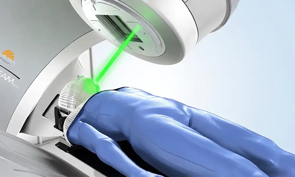
Treatment process
1. MDT: Comprehensive assessment of the patient's condition
Assess suitability for radiotherapy by evaluating contrast-enhanced CT/MRI scans, organ function, and prior treatment history; define radiotherapy goals; and seamlessly integrate radiotherapy with other treatment modalities such as surgery, targeted therapy, and immunotherapy.
2. Personalized patient immobilization + 4D localization scanning
Fabricate a custom immobilization mold and perform 4D-CT localization; for lesions with significant respiratory motion, capture full-cycle respiratory images to provide comprehensive data for treatment planning.
3. Dual-person review of target volume delineation and treatment planning
Radiation oncologists delineate the tumor target area and organs at risk, while medical physicists optimize beam angles and fractional doses;the treatment plan is executed only after being double-checked and approved by two professionals.
4. Implementation of fractionated precision radiotherapy
① CBCT 3D image-guided positioning correction;
② 6-degree-of-freedom (6DoF) robotic couch for automatic micro-adjustment of patient position;
③ RapidArc volumetric modulated arc therapy (VMAT);the entire process is non-invasive, and most patients can complete treatment on an outpatient basis.
5. Standardized, stratified follow-up and re-examination
Conducts scheduled follow-ups involving imaging and blood tests to monitor lesion dynamics and reactions to radiotherapy, adjusting supportive and systemic treatment regimens as necessary.
Safety and Precautions
1. MDT: Comprehensive preoperative assessment as the foundation for safety
Comprehensively assess tumor burden, the distance between lesions and sensitive organs, and the tolerance of the heart, lungs, liver, and kidneys; anticipate potential adverse reactions and formulate a tailored fractionated dosage plan.
2. Continuous image tracking reduces radiation targeting errors.
Real-time monitoring of the target area's position is achieved using CBCT and 4D respiratory gating, reducing unnecessary radiation exposure to normal tissues.
3. Short-term, transient, mild reactions
Some patients may experience mild redness in the treated area, transient fatigue, or loss of appetite. These are generally reversible, short-term effects that gradually subside with proper care as advised by your doctor.
4. Specialized skin care for the irradiated area
During radiotherapy and for several months afterward, the treated area must not be exposed to direct sunlight, friction, hot compresses, or irritating cleansing/skincare products;seek a follow-up consultation immediately if skin breakdown or persistent pain occurs.
5. Prompt medical consultation and assessment for abnormal symptoms
If symptoms such as persistent high fever, severe nausea and vomiting, limb numbness, severe skin damage, or persistent pain upon swallowing occur, contact medical staff immediately for medical attention.
6. Unified planning of combination therapy by the multidisciplinary team
Vital Beam radiotherapy can be combined with interventional therapy, ablation, targeted therapy, and immunotherapy; the MDT (Multidisciplinary Team) coordinates the treatment intervals, medication cycles, and follow-up schedules.
FAQs
1. What is the VitalBeam linear accelerator?
VitalBeam is a medical linear accelerator radiotherapy system that delivers external radiation therapy to tumor target areas by generating high-energy X-rays or electron beams.It supports a range of precision radiotherapy functions, depending on the specific configuration chosen by the hospital.
2. What radiotherapy techniques can be performed using VitalBeam?
Depending on equipment configuration and hospital qualifications, available treatments include 3D conformal radiotherapy, intensity-modulated radiotherapy (IMRT), volumetric modulated arc therapy (VMAT), image-guided radiotherapy (IGRT), and certain forms of stereotactic therapy.Actual availability of features such as respiratory gating depends on the specific equipment configuration at the facility.
3. What is the difference between VitalBeam and CyberKnife?
VitalBeam utilizes a gantry-based linear accelerator design, making it suitable for a wide range of needs, from conventional fractionation to various forms of precision radiotherapy. In contrast, the CyberKnife system employs a robotic arm, excelling in non-coplanar, multi-angle irradiation and motion tracking.Both systems are capable of delivering precision radiotherapy, and the choice between them should be based on the specific characteristics of the lesion.
4. Which tumors can be treated with VitalBeam?
Tumors assessed by a radiation oncologist as requiring external beam radiotherapy may be considered for treatment; this includes sites such as the head and neck, thorax, abdomen, pelvis, bone, and soft tissue.Specific techniques and dosages depend on the pathological type, stage, and treatment objectives.
5. Why are localization and mold/cast preparation required before treatment?
Localization CT is used to determine the spatial positions of the lesion and normal organs, while immobilization devices help the patient maintain a consistent posture during each treatment session.Physicians and medical physicists design and review the dose distribution to confirm target coverage and the protection of normal organs.
6. How long does a single treatment session take?What are the potential side effects?
The actual beam delivery time is usually short; however, when patient positioning and image verification are included, the entire process may take longer.Side effects—such as fatigue, skin reactions, throat discomfort, or gastrointestinal reactions—depend on the treatment site and cumulative dose; it cannot be assumed that all patients will experience only mild reactions.


