


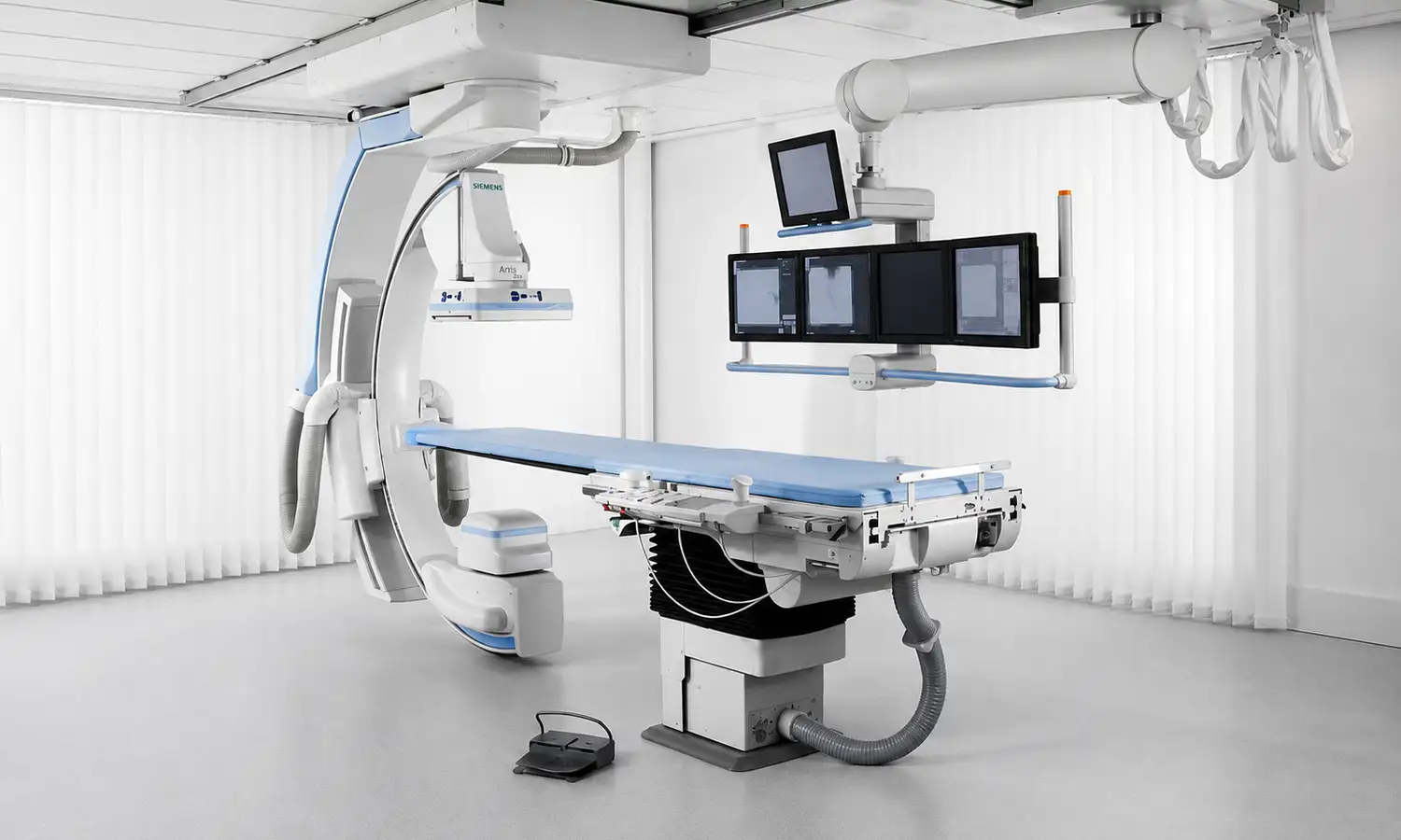
Developed by Siemens Healthineers (Germany), the Artis zee Ceiling is a classic ceiling-mounted model within the Artis zee series.It utilizes digital subtraction technology to remove images of background tissues—such as bone and muscle—while retaining only the pure vascular image, thereby providing clinicians with a clear and precise "map" of the blood vessels.The system utilizes an 11×14-inch large flat-panel detector and is compatible with both dedicated small-format cardiac detectors and general-purpose large-format detectors;it features a ceiling-mounted robotic arm design.
Core Technologies
What is DSA?
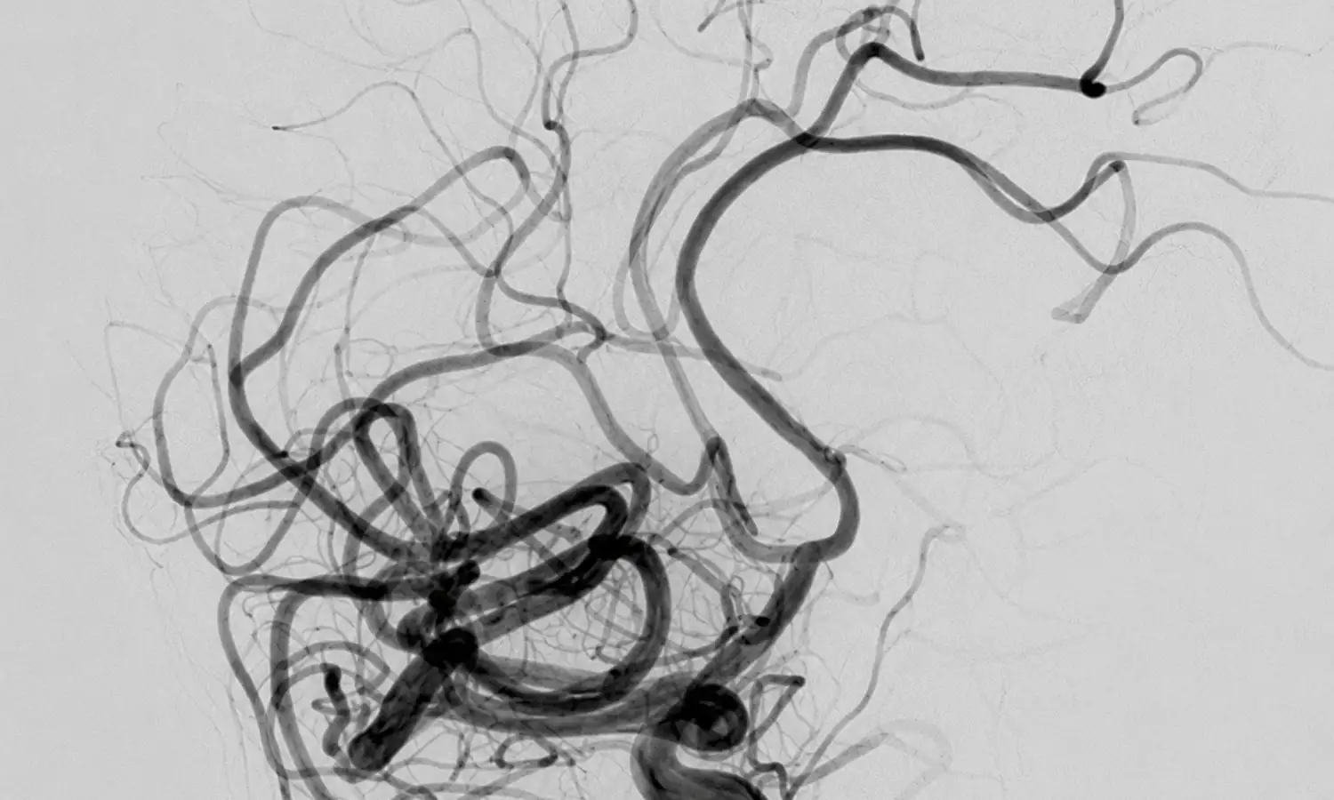
DSA is an advanced X-ray imaging system that combines conventional angiography with computer image processing technology.During the procedure, a physician inserts a catheter into the target blood vessel and injects a contrast agent, while the equipment rapidly captures a series of X-ray images.Using digital subtraction technology, the system automatically removes background images—such as bone and muscle—leaving only a clear image of the blood vessels.This provides the physician with real-time dynamic images of the vasculature, clearly displaying abnormalities such as stenosis, occlusion, aneurysms, and malformations.
1. High-definition digital flat-panel detector and rapid imaging technology
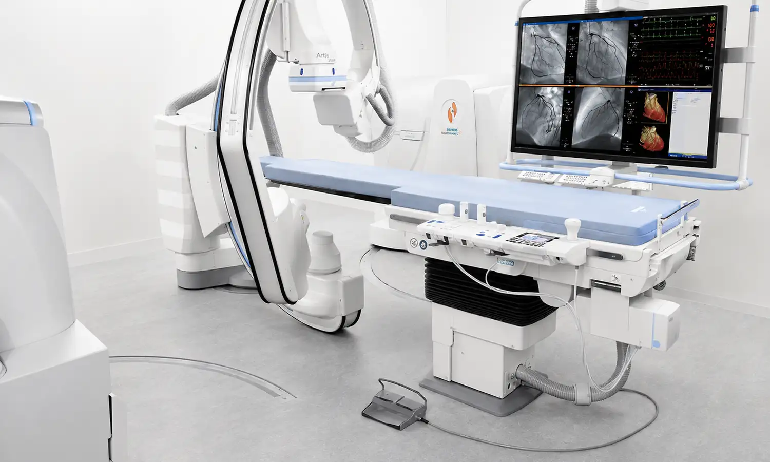
The Artis zee ceiling utilizes a high-resolution digital flat-panel detector to effectively minimize image distortion while enhancing both spatial and density resolution. Its imaging dynamic range is ten times that of conventional DSA systems.By employing intelligent noise reduction and adaptive vascular edge enhancement technologies, the system produces clearer images characterized by sharp vascular edges and refined background detail.The advanced ceiling-mounted gantry and the bezel-free design of the flat-panel detector facilitate comprehensive patient access and rapid gantry positioning.The C-arm supports a maximum rotation speed of 60 degrees per second.
2. Intelligent low-dose and 3D imaging technology
The X-ray dose required for image acquisition with the Artis zee ceiling system is approximately 50% of that used in conventional DSA. Thanks to Siemens' proprietary fluoroscopy dose regulation and fluoroscopy storage technologies, physicians can obtain clear, diagnostic-quality stored fluoroscopy images without being exposed to high radiation doses in the procedure room.Equipped with DynaCT functionality, the system enables the direct acquisition of CT-like image data during interventional procedures, eliminating the need to transfer patients for separate CT or MRI scans and facilitating precise positioning for interventions such as punctures, drainage, and biopsies.Additionally, the system features stepping DSA angiography for lower-extremity vessels, allowing for full-length vascular imaging in a single pass; rotational DSA for viewing vasculature from multiple angles; and 3D reconstruction capabilities supporting various methods, including Shaded Surface Display (SSD), Maximum Intensity Projection (MIP), Volume Rendering (VR), and Multi-Planar Reconstruction (MPR).
Scope of Indications
The Artis zee Ceiling digital flat-panel DSA is widely used for the diagnosis and interventional treatment of vascular diseases throughout the body.
1. Major clinical applications
Field Primary Applications
Cardiovascular intervention Coronary angiography (CAG), percutaneous coronary intervention (PCI), radiofrequency ablation for arrhythmia, congenital heart disease closure, pacemaker implantation, etc.
Neuro-interventional procedures: cerebral angiography, intracranial aneurysm embolization, emergency thrombectomy for cerebral infarction, arteriovenous malformation embolization, etc.
Peripheral vascular intervention (e.g., balloon dilation and stent implantation for peripheral arterial stenosis/occlusion, lower-extremity arterial intervention)
Interventional Oncology: Includes various tumor embolization procedures, perfusion chemotherapy, and the CLEARmap one-stop interventional oncology service.
Comprehensive interventional procedures Percutaneous biopsy, drainage, emergency interventional hemostasis for bleeding, etc.
2. Suitable patient population
• Individuals requiring cardiovascular or cerebrovascular interventional diagnosis and treatment
For patients requiring interventional procedures for conditions such as coronary artery disease, cerebrovascular stenosis, or intracranial aneurysms, the Artis zee ceiling system ensures precise intraoperative image guidance.
• Individuals requiring interventional tumor therapy
For patients with solid tumors (e.g., liver cancer) requiring transarterial chemoembolization (TACE), the equipment precisely visualizes tumor-feeding vessels, guiding the catheter for super-selective cannulation.
• Patients with peripheral vascular disease and emergency hemorrhage
For patients with peripheral artery stenosis, lower-extremity arterial occlusion, or various forms of acute hemorrhage, the equipment can rapidly identify the bleeding site and guide interventional hemostasis.
Core Strengths
Comparison Dimension Conventional Angiography Artis zee Ceiling Digital Flat-Panel DSA
Imaging quality Significant background interference (bones, muscles, etc.) Digital subtraction technology; clear vascular images
Radiation dose Relatively high Dose reduced by approx. 50%; eco-friendly and safe
Imaging dynamic range Limited 10 times that of conventional DSA
3D imaging Not achievable DynaCT (CT-like imaging) allows for the direct acquisition of 3D information during surgery.
Contrast agent dosage: Significantly reduced, lowering the risk of renal impairment.
Catheter/Guidewire Visibility: Limited clarity; however, low-density catheters, guidewires, and stents are clearly visualized.
Gantry flexibility: Limited vs. Suspended design with 60°/sec rotation for omnidirectional coverage.
Conventional Angiography
While conventional angiography visualizes vascular structures, background images—such as bone and soft tissue—can interfere with vascular observation during complex interventional procedures, potentially hindering the precise assessment of lesions.Limitations in dynamic range and spatial resolution capabilities restrict the visualization of fine vascular structures and low-density interventional devices.Furthermore, conventional equipment faces limitations regarding 3D imaging, catheter navigation, and real-time intraoperative assessment.
Artis zee Ceiling Digital Flat-Panel DSA
The Artis zee ceiling digital flat-panel DSA system employs digital subtraction technology to effectively eliminate background interference from bone and soft tissue, delivering clearer vascular images that assist physicians in precisely identifying vascular lesions and interventional pathways.With an enhanced dynamic range, the system improves the visualization of complex vascular structures and minute lesions.During interventional procedures, the Artis zee ceiling supports 3D image acquisition, enabling the intraoperative capture of CT-like 3D vascular data to inform precise localization and treatment planning.Additionally, the system clearly visualizes low-density interventional devices—such as catheters, guidewires, and stents—thereby enhancing procedural accuracy.The system also optimizes radiation control through low-dose imaging technology and reduces contrast agent usage, alleviating the burden on patients during examinations and treatments.Its ceiling-mounted gantry design, combined with high-speed rotation capabilities, ensures flexible, multi-angle coverage, providing stable and efficient imaging support for complex interventional surgeries.
Clinical Application Case
1. Interventional treatment for recurrent aneurysms
The patient had previously undergone interventional treatment for a ruptured anterior communicating artery aneurysm, but follow-up examinations revealed a recurrence.Imaging showed an irregularly shaped aneurysm with a risk of re-rupture.Due to the complex location of the aneurysm, traditional interventional embolization posed challenges—such as technical difficulty and recurrence risk—necessitating precise assessment of the vascular structure and the formulation of a personalized treatment plan.
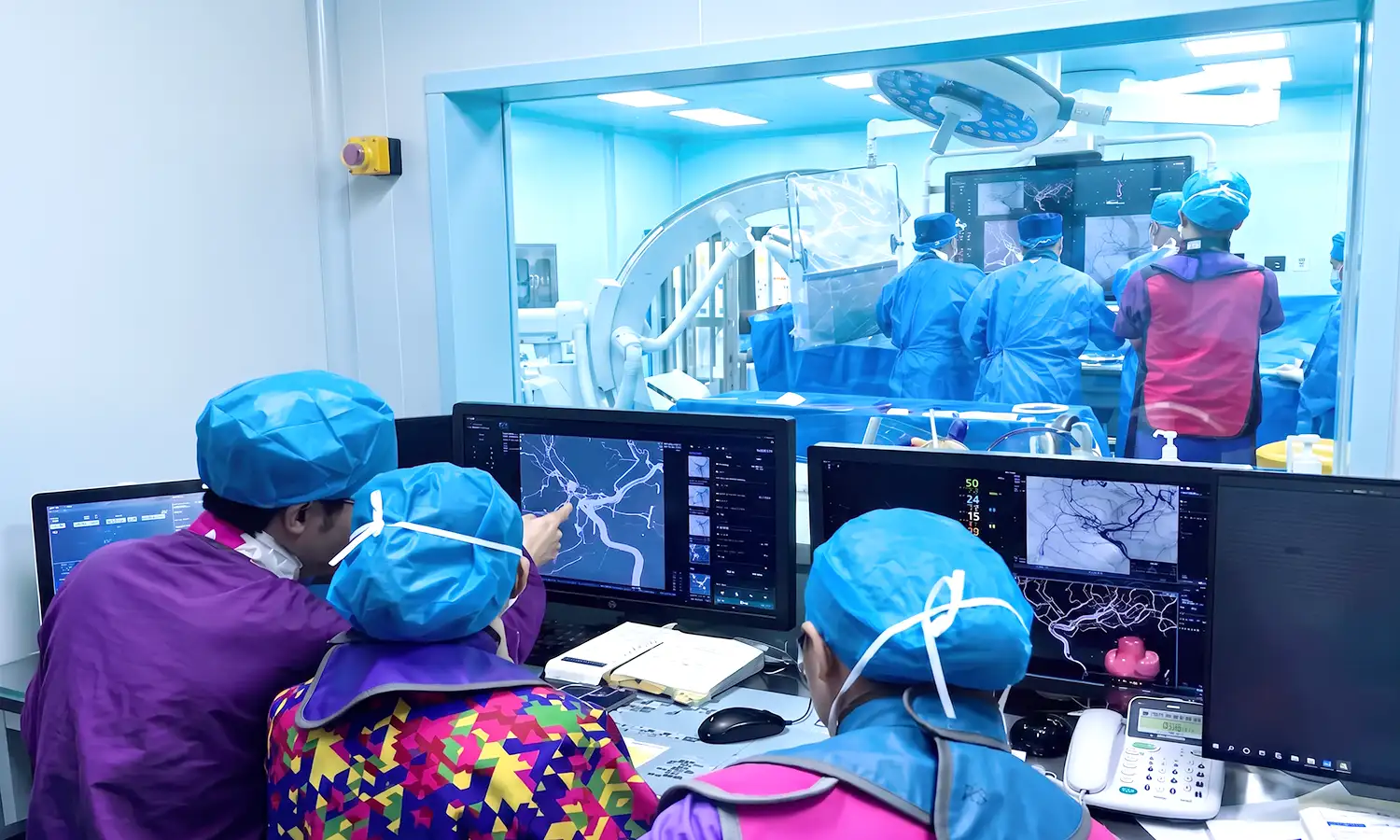
To enhance treatment precision, the medical team utilizes the German Siemens Artis zee ceiling digital flat-panel DSA system to assist in neurointerventional procedures.Preoperative DSA angiography clearly reveals the aneurysm's location, size, and relationship with surrounding blood vessels, providing precise imaging data for surgical planning.
• Treatment regimen
DSA-guided embolization of intracranial aneurysms using the WEB device
• Outcomes and follow-up
During the procedure, the DSA system provided real-time visualization of vascular structures and the position of interventional instruments, facilitating the precise deployment of the WEB device.Postoperative angiography showed no residual aneurysm filling and confirmed the patency of the parent artery.The patient recovered well and successfully completed rehabilitation.
FAQ
1. Is DSA a diagnostic procedure or a treatment?
DSA can be used for angiographic examinations and provides real-time image guidance for endovascular interventional procedures.The decision to perform diagnostic imaging alone or combined treatment is made by the physician based on the patient's condition, imaging results, and the pre-procedural plan.
2. Does DSA require surgery or general anesthesia?
Most angiography and interventional procedures involve accessing blood vessels via percutaneous puncture, eliminating the need for large traditional incisions; some procedures can be performed under local anesthesia.Patients undergoing complex procedures or those unable to cooperate may require sedation or general anesthesia, subject to assessment by the anesthesia and interventional teams.
3. Why is a contrast agent used during a DSA procedure?
Iodine-based contrast agents allow blood vessels to appear clearly on X-ray images, helping doctors identify conditions such as stenosis, blockages, hemorrhages, or vascular abnormalities.Patients with a history of adverse reactions to contrast agents, abnormal renal function, thyroid disease, or relevant medication use should inform the doctor in advance.
4. What preparations are required before a DSA procedure?
Routine tests—such as complete blood count (CBC), coagulation profile, and liver/kidney function tests—are typically required.Patients must fast (abstain from food and drink) or adjust their intake of anticoagulants, antiplatelet agents, and hypoglycemic medications as instructed by the physician.Patients should not discontinue medications on their own but must truthfully inform the medical team about all current medications and any allergies.
5. How do I schedule a DSA examination or inquire about interventional therapy?
If you require a DSA examination or wish to learn about interventional therapy, please prepare recent imaging scans, relevant laboratory test results, and records of past surgeries and medications so that an interventional radiologist can assess the indications for the procedure.When scheduling an appointment, please proactively disclose any history of adverse reactions to contrast agents, your renal function status, and the use of anticoagulants or antiplatelet drugs.