


Indications
1. Applicable conditions
• Solitary lesions ≤3 cm in diameter where complete local ablation is anticipated;lesions 3–5 cm in diameter may be treated using a staged approach combined with TACE
• Elderly patients with multiple underlying conditions and poor cardiopulmonary or hepatic/renal reserve who cannot tolerate open surgical resection.
• Postoperative patients with local residual disease or oligorecurrent solid lesions requiring repeat local intervention
• Patients who wish to preserve organ shape and physiological function and prefer minimally invasive treatments.
• Intermediate-to-advanced stage disease with oligometastases; used as a local adjunctive measure for tumor debulking and symptom relief.
2. Common indications
Localized solid lesions such as primary liver cancer, liver metastases, peripheral lung cancer, lung metastases, thyroid nodules/low-risk thyroid cancer, renal tumors, small breast nodules, prostate lesions, uterine fibroids, and bone metastases.
Core Strengths
1. Rapid temperature rise;higher ablation efficiency
High-frequency electromagnetic waves directly excite tissue molecules to generate heat, rapidly reaching the temperature required for effective inactivation.This shortens the procedure time for individual lesions, thereby reducing patient discomfort associated with maintaining a fixed position.
2. Resistant to heat-sink effects from blood flow; suitable for highly vascularized lesions
Compared to other thermal ablation methods, the microwave thermal field is less affected by heat dissipation caused by blood flow; this allows for the formation of a stable ablation zone even in tumors located adjacent to major blood vessels or those with a rich blood supply.
3. Minimally invasive puncture to maximize preservation of organ function
Only a tiny wound from the needle tract remains, without the need for extensive tissue resection; it is a preferred choice for areas where appearance and function are critical, such as the thyroid and breast, reducing the risk of postoperative functional impairment.
4. Supports multiple repeat ablation sessions
Provided that organ function tolerance allows, the procedure can be repeated in fractions for new or residual lesions, offering a repeatable strategy for long-term lesion control.
5. Flexible synergistic combination of multiple regimens
TACE (Transarterial Chemoembolization) can be performed first to shrink the tumor prior to ablation; it can also be combined synchronously or sequentially with targeted therapy, immunotherapy, and radiotherapy to create a comprehensive local-systemic integrated strategy.
Technical features
1. MDT: Upfront design of personalized treatment plans
Joint consultation involving interventional radiologists, oncologists, radiologists, and surgeons; ablation power and puncture paths are selected based on lesion size, proximity to blood vessels, and organ function, determining whether to use standalone ablation or a combined interventional approach.
2. Precision navigation using multimodal imaging
Equipped with a fusion positioning system combining ultrasound and contrast-enhanced CT, enabling real-time intraoperative monitoring of ablation margins and precise thermal control for lesions near the trachea, bile ducts, and major blood vessels, thereby minimizing damage to normal tissue.
3. Standardized quality control system covering the entire process
Strictly adhere to domestic expert consensus on microwave ablation; conduct comprehensive pre-operative screening of coagulation status and organ function; dynamically adjust energy levels during the procedure;and perform immediate post-ablation imaging to assess the coverage area.
4. One-stop comprehensive diagnosis and treatment coordination
The ablation center is integrated with interventional radiology, medical oncology, and radiation oncology departments; treatment schedules for ablation, interventional procedures, and systemic therapies are coordinated to minimize the need for patients to travel between multiple departments.
5. Stratified, long-term follow-up management
Scheduled contrast-enhanced imaging follow-ups at 1, 3, and 6 months post-procedure to dynamically monitor lesion changes and, as needed, perform supplementary ablation or adjust the systemic anti-tumor regimen.
Technical definition
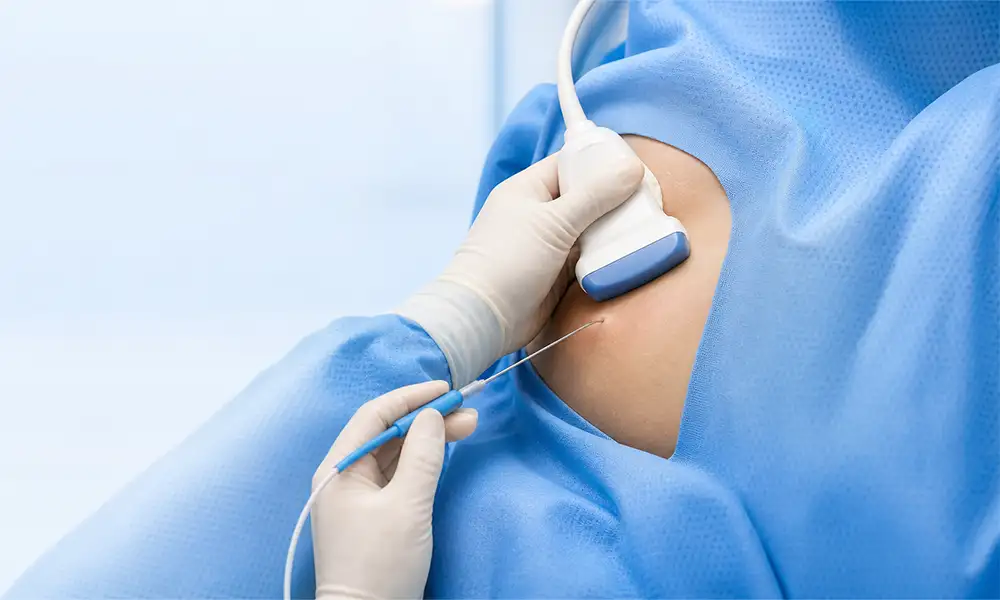
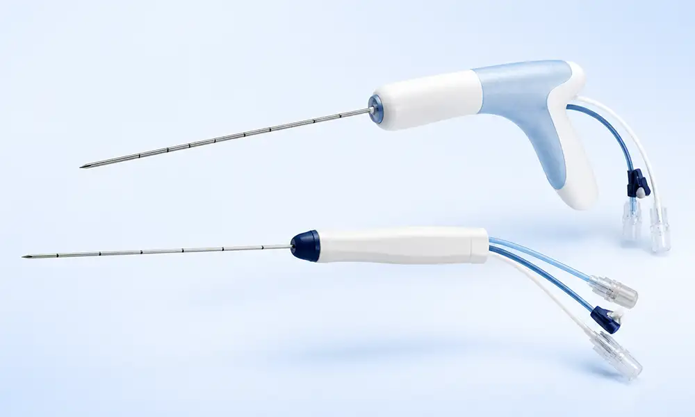
Microwave ablation (MWA) is an image-guided, minimally invasive thermal ablation technique. Under real-time CT or ultrasound monitoring, a microwave antenna is inserted into the tumor; high-frequency electromagnetic waves cause rapid friction among polar molecules in the tissue, generating heat that raises the local temperature to a stable 60–150°C, thereby inducing coagulative necrosis of the tumor.Procedures follow the national expert consensus on microwave ablation for solid tumors, making it a key local intervention within multidisciplinary comprehensive treatment.
Working principle/mechanism
The microwave generator emits high-frequency electromagnetic waves. When the antenna acts upon the lesion, polar molecules—such as water and proteins—vibrate and collide at high speeds, rapidly converting energy into heat. The entire lesion reaches an inactivation temperature within a short period, destroying cell membranes and causing irreversible protein denaturation.Microwave energy creates a uniform thermal field; because the cooling effect of blood flow is minimal, a stable ablation zone is maintained even when the lesion is near blood vessels. Simultaneously, the procedure seals the lesion's tiny blood supply vessels, doubly inhibiting lesion progression, while real-time imaging monitors the thermal boundaries throughout the process.
Treatment process
1. Comprehensive preoperative assessment: Complete contrast-enhanced CT/MRI, liver and kidney function tests, and coagulation studies; conduct an MDT consultation to determine suitability for ablation and formulate a single-needle or multi-needle ablation plan.
2. Image-guided localization and marking: Ultrasound or CT scans mark the skin entry point and calculate a safe needle path, avoiding nerves, blood vessels, and hollow organs.
3. Puncture under local anesthesia: A microwave antenna is precisely placed into the tumor target area via percutaneous puncture under sedation and analgesia.
4. Segmented microwave thermal ablation: Power and ablation duration are set based on lesion size, with real-time intraoperative monitoring of the thermal field.
5. Needle tract cauterization for hemostasis: Upon completion of ablation, the needle is withdrawn while simultaneously cauterizing the puncture tract to reduce the risk of bleeding and tumor seeding along the tract;pressure-bandaging is applied.
6. Scheduled imaging follow-up: Periodic post-operative contrast-enhanced scans to assess the inactivation status of the lesion and local changes.
Safety and Precautions
1. MDT: Preoperative screening as the foundation for safety
Strictly screen for and exclude patients unsuitable for ablation—such as those with diffuse multiple tumors, severe organ failure, serious coagulation disorders, or active infections—and anticipate the risk of thermal injury in advance.
2. Continuous dynamic imaging monitoring throughout the procedure
Real-time observation of the expansion of the ablation heat zone; energy output per session is reduced when treating areas adjacent to critical organs, and ablation is performed in multiple zones and fractions.
3. Transient normal post-operative reactions
Some patients may experience a brief low-grade fever, dull pain at the puncture site, or mild fatigue. These are usually stress reactions related to tissue absorption following ablation and will gradually resolve with appropriate symptomatic treatment.
4. Specialized care for the puncture site
Keep the post-procedure puncture site dry and clean; avoid lifting heavy objects or strenuous exercise for 1–2 weeks to prevent hematoma or oozing.
5. Prompt medical consultation for abnormal symptoms
If symptoms such as persistent high fever, severe chest or abdominal pain, hemoptysis (coughing up blood), worsening jaundice, or difficulty breathing occur, contact medical staff immediately for assessment.
6. Unified coordination of comprehensive treatment
The timing intervals and medication schedules for combining ablation with TACE, targeted therapy, or immunotherapy are determined by a multidisciplinary team; follow-ups and treatments must not be interrupted without medical advice.
FAQs
1. What is microwave ablation?
Microwave ablation generates microwave energy via an ablation needle inserted into the lesion, causing water molecules within the tissue to vibrate and rapidly heat up, thereby inducing coagulative necrosis.
2. Which lesions might be suitable for microwave ablation?
Suitable for selected solid tumors (e.g., liver, lung, kidney) and certain benign nodules (e.g., thyroid, breast).Suitability depends on pathological characteristics, size, number, location, and the patient's overall condition.
3. What are the characteristics of microwave ablation compared to radiofrequency ablation?
Microwaves typically generate heat rapidly, creating a relatively large ablation zone, and are often less affected by heat dissipation from blood flow compared to other methods.However, this does not mean microwave ablation is superior to radiofrequency ablation for all lesions; equipment, tumor location, and physician experience are equally important factors.
4. Does microwave ablation affect the organ's original function?
The treatment goal is to cover the lesion while preserving normal tissue as much as possible; however, complete avoidance of impact on organ function cannot be guaranteed.When the lesion is close to the bile duct, airway, nerves, or other vital structures, protective measures—such as isolation or cooling—must be implemented, or an alternative treatment plan selected.
5. How soon can one recover after treatment?
Recovery time depends on the treatment site, ablation scope, anesthesia method, and presence of complications.Most patients can gradually resume light activity but should avoid strenuous exercise, heavy lifting, and straining the puncture site in the short term.
6. Under what circumstances might a repeat ablation be necessary?
If follow-up reveals active lesions at the ablation margin, or if new localized lesions appear subsequently, repeat treatment may be considered.The final decision should be based on contrast-enhanced imaging, organ function, and the overall tumor control plan.


