


Indications
1. Applicable conditions
• Lesions immediately adjacent to vital structures such as major blood vessels, nerves, or the chest wall, requiring strict safety margins
• Patients with tumors in challenging locations or multiple underlying conditions who cannot tolerate general anesthesia or surgical resection.
• Postoperative patients with local recurrence lesions requiring repeat local intervention and optimization of the comprehensive treatment plan
• Elderly patients with multiple underlying conditions who seek minimally invasive treatment, short recovery periods, and preservation of organ function
2. Common indications
• Malignant solid tumors (e.g., liver cancer, lung cancer, kidney cancer, prostate cancer, bone and soft tissue tumors); suitable for cases requiring organ function preservation or where surgery is not an option.
• Benign lesions: Breast fibroadenomas, uterine fibroids, and other benign space-occupying lesions
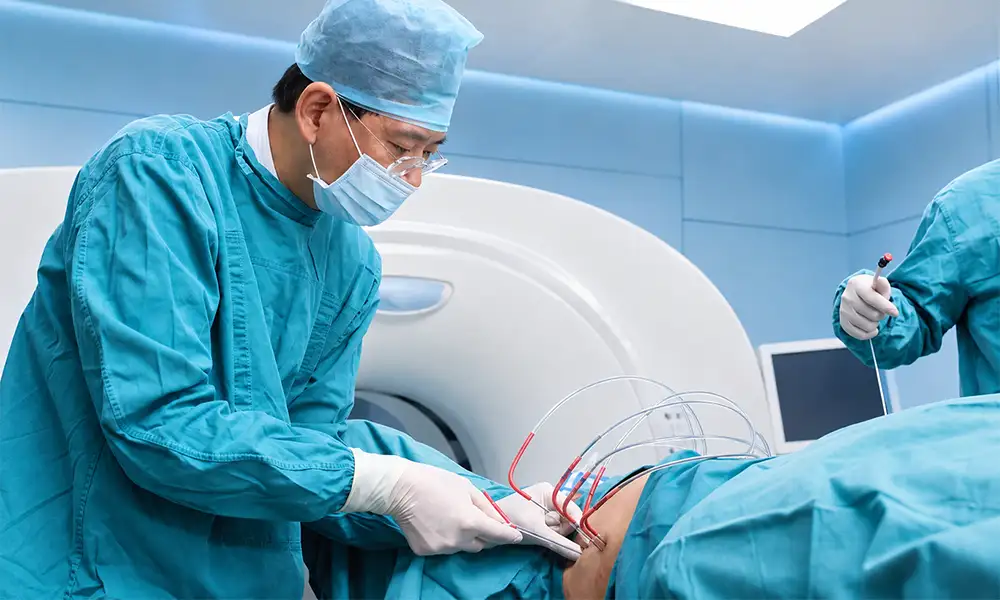
Core Strengths
Cryoablation is a distinctive technique within the field of minimally invasive tumor ablation. It offers a gentle, controllable local treatment option—determined after multidisciplinary assessment—for complex lesions where traditional surgery or thermal ablation carries higher risks.
1. Ice ball fully visualized throughout the procedure; boundaries precisely controllable
The low-temperature ice ball formed during cryoablation is visible on imaging in real-time, allowing for intuitive intraoperative assessment of the ablation coverage area and enhancing procedural control.
2. Gentler on blood vessels and nerves
Cryogenic effects cause minimal irritation to adjacent major blood vessels and nerves; provided there is no direct puncture-related injury, the risk of damage to vital structures is reduced.
3. Inherent analgesic effect; minimal pain during and after the procedure
Extreme cold can induce a local anesthetic effect; consequently, most patients experience relatively mild pain during and after the procedure.
4. Minimally invasive puncture; short recovery period
The procedure is performed through a tiny percutaneous puncture tract; no laparotomy or thoracotomy is required; hospital stays are shortened, and physical trauma is minimized.
5. Broad applicability; suitable for complex cases
Addresses benign and malignant lesions and recurrences across multiple organs, as well as cases involving elderly or frail patients, providing local intervention pathways for those unable to tolerate surgery.
Technical features
We utilize a comprehensive clinical cryoablation system covering lesions in the liver, lung, kidney, bone, breast, and prostate.Equipped with a complete argon-helium cryoablation system and high-definition CT image guidance, the procedure allows for real-time visualization and boundary control of the "ice ball," enhancing operational safety.The ablation team is led by chief physicians with extensive experience in minimally invasive interventional procedures and strictly adheres to a multidisciplinary team (MDT) consultation protocol: pre-operative planning involves collaboration among oncology, radiology, and surgery departments to determine puncture paths and freeze-thaw strategies; post-operative care includes regular imaging follow-ups to dynamically assess lesion changes and coordinate with targeted therapy, immunotherapy, and radiotherapy to formulate an integrated, comprehensive treatment plan.The hospital continuously tracks evidence-based research on cryoablation and optimizes personalized procedural workflows for tumors in different anatomical locations.
Technical definition
Cryoablation is an image-guided, minimally invasive local ablation technique. It delivers high-pressure argon and helium gases through a puncture probe to subject the lesion area to repeated cycles of ultra-low-temperature freezing and rapid rewarming. This process induces irreversible damage to tumor cells, thereby achieving local control of the lesion. The procedure requires only local anesthesia and involves a minimal incision, making it a key method in the comprehensive local treatment of solid tumors.Cryoablation is a minimally invasive therapy that destroys diseased tissue using extreme cold.By delivering argon and helium gases to the target area via a cryoprobe, local tissue undergoes repeated cycles of extreme freezing and rapid rewarming within a short period, causing irreversible damage and loss of viability in tumor cells.
Working principle/mechanism
When the argon-helium cryoprobe releases argon gas, the tip temperature rapidly drops to between -140°C and -170°C, creating a cryogenic ice ball that encapsulates the lesion; switching to helium gas allows for rapid rewarming, and the freeze-thaw cycle is typically repeated two to three times.
During the freeze-thaw process, the repeated formation and melting of intracellular ice crystals directly damage cell membranes and internal structures. Simultaneously, the low temperature causes the occlusion of microvessels within the lesion, cutting off the tumor's nutrient supply; this dual action leads to the inactivation and necrosis of the targeted tumor tissue.Real-time imaging monitoring throughout the procedure ensures precise control over the cryogenic zone, minimizing impact on surrounding healthy tissue.
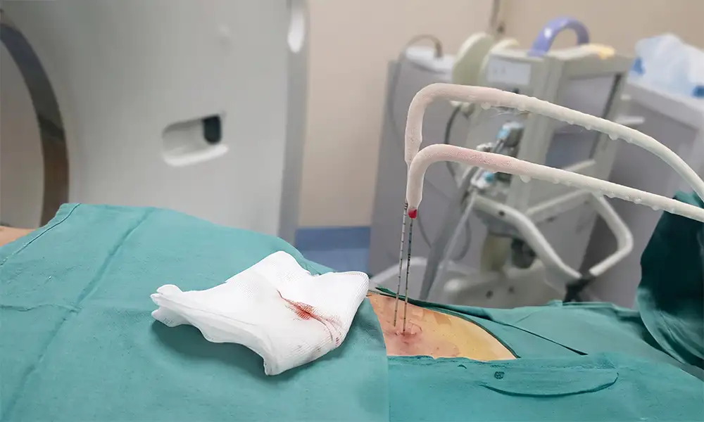
Treatment process
1. Comprehensive preoperative assessment: Complete contrast-enhanced CT/MRI, CBC, coagulation studies, and organ function tests; the MDT team evaluates the spatial relationship between the lesion and surrounding organs to determine the puncture and freeze-thaw protocol.
2. Precision image-guided localization: CT, ultrasound, or MRI scans pinpoint the lesion and plan a safe puncture path.
3. Precise probe placement: Following local anesthesia, a cryoprobe is delivered to the tumor target area via percutaneous puncture.
4. Standardized freeze-thaw cycles: Cryogenic freezing is maintained for a set duration, followed by rewarming with helium gas; this standard cycle is repeated 2–3 times.
5. Probe removal and wound management: Remove the probe after confirming that the ice ball completely covers the target lesion;apply pressure and a dressing to the puncture site.
6. Long-term imaging follow-up: Periodic post-operative contrast-enhanced imaging to assess lesion changes and simultaneously adjust the subsequent comprehensive treatment plan.
Safety and Precautions
The safety of cryoablation relies primarily on real-time monitoring via imaging equipment to precisely control the zone of cryogenic action.Before the procedure, physicians carefully analyze the spatial relationship between the lesion and surrounding blood vessels, nerves, and organs to plan a safe puncture path.Post-procedure, a small number of patients may experience transient local swelling, pain, or mild fever; such discomfort usually resolves on its own.A robust pre-operative assessment system and standardized procedures performed by experienced physicians significantly reduce the likelihood of complications.
FAQs
1. What is cryoablation?How are tumors "frozen to death"?
Cryoablation involves inserting a cryoprobe into the lesion under CT, ultrasound, or MRI guidance. Through cycles of freezing and thawing, ice crystals form both intracellularly and extracellularly, disrupting cell membranes and microcirculation, thereby causing the loss of viability in the targeted tissue.
2. Which tumors and benign lesions are suitable for treatment via cryoablation?
Suitable for selected localized lesions in the liver, lung, kidney, prostate, bone, and soft tissue, as well as certain benign lesions.Suitability depends primarily on lesion size, number, location, proximity to critical surrounding structures, and the patient's overall physical condition.
3. What are the differences between cryoablation, radiofrequency ablation, and microwave ablation?
RF and microwave ablation destroy lesions by raising the temperature, whereas cryoablation works through freezing and thawing cycles.The "ice ball" formed during cryoablation is usually visible on imaging and offers some local analgesic effects; however, each of the three technologies has its own specific applications, and no single method is superior for every type of lesion.
4. Is anesthesia required during the treatment process?Will there be any pain?
Depending on the location of the lesion and the complexity of the puncture, local anesthesia, sedation, or general anesthesia may be used.While low temperatures provide some analgesic effect, the puncture, tract dilation, and post-procedural inflammation may still cause aching or pain.
5. How many cryoablation sessions are needed?How long does recovery take?
Small, localized lesions may be treated in a single session, whereas larger, multiple, or complexly located lesions may require staged treatment.Recovery time varies depending on the treatment site and the patient's physical condition; most patients can resume light activity relatively soon, but specific instructions provided by the medical team upon discharge should be followed.
6. What are the potential risks?How is treatment efficacy assessed?
Risks include hemorrhage, infection, injury to adjacent tissues, and pneumothorax (following pulmonary treatment); while severe complications are relatively rare, they cannot be entirely ruled out.Post-procedure assessment—typically via contrast-enhanced CT, MRI, or ultrasound—is required to verify that the ablation zone fully covers the lesion;assessment based solely on symptoms is insufficient.


