


Skin cancer is the most common malignant tumor globally, with basal cell carcinoma accounting for 80% of cases and squamous cell carcinoma for 20%.It frequently occurs on sun-exposed areas (such as the head, face, and backs of the hands).Solar UV radiation is the primary risk factor.Early detection and surgical excision result in a cure rate exceeding 95%.
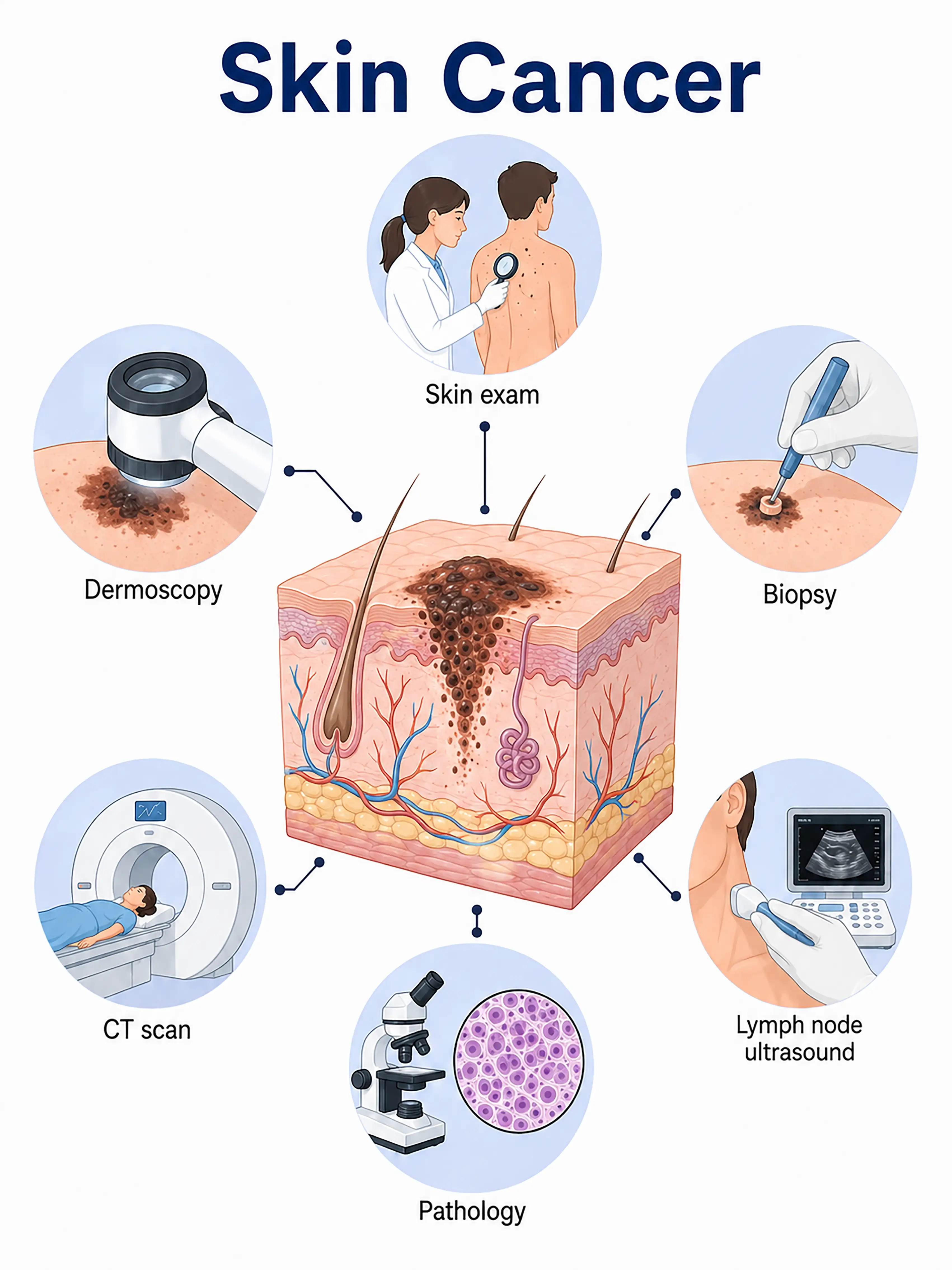
Diagnosis based on skin cancer stage
1.Early diagnosis of skin cancer
A new skin growth appears, or an existing mole undergoes changes.Basal cell carcinoma presents as pearly papules or ulcers, while squamous cell carcinoma appears as keratotic plaques or nodules.Diagnosis is confirmed via dermoscopic observation of characteristic structures and excisional biopsy.
2.Intermediate-stage skin cancer diagnosis
The lesion enlarges, presenting with ulceration, bleeding, or pain.Biopsy determines the pathological type and degree of differentiation, and the depth of invasion into surrounding tissues is assessed.
3.Advanced skin cancer diagnosis
Tumor extensively invades deep tissues or metastasizes to distant lymph nodes and viscera.Whole-body CT and PET-CT are used to assess the extent of metastasis.
If not diagnosed promptly, skin cancer—while rarely metastasizing—can cause extensive local tissue destruction; facial tumors, in particular, can severely affect appearance and function.
When to Seek Medical Attention
Early signs of skin cancer include new growths on the skin, ulcers that do not heal, or changes to existing moles.Please consult a doctor via "Chat with a Doctor" for early detection.During the online consultation, the doctor will ask about your history of sun exposure and skin changes, and provide guidance on undergoing a skin biopsy.
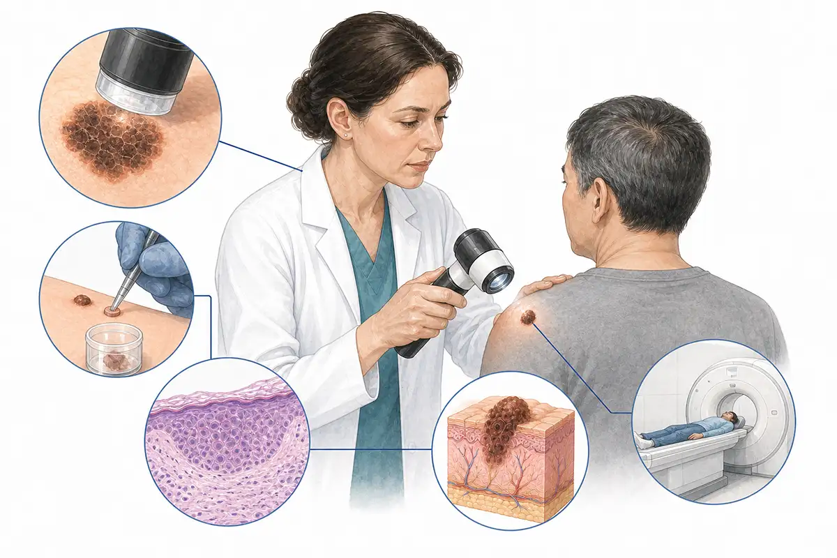
Basis for Skin Cancer Diagnosis
1.Clinical skin examination
Comprehensive visual inspection and palpation of all skin areas—observing the morphology, color, borders, and surface characteristics of lesions—serve as the fundamental method for initial screening.
2.Dermoscopy
Dermoscopy allows for the magnified observation of skin lesions; basal cell carcinoma may show arborizing vessels and blue-gray ovoid nests, while squamous cell carcinoma may show keratin plugs and atypical vessels.
3.Resection or incisional biopsy
Obtaining lesional tissue for pathological examination is the gold standard for diagnosing skin cancer; it clarifies the pathological type, degree of differentiation, and depth of invasion.
4.Curettage biopsy
Curettage biopsy is performed on superficial lesions to rapidly obtain tissue samples for pathological diagnosis; this method is suitable for suspected superficial basal cell carcinoma.
5.Skin ultrasound
High-frequency ultrasound measures tumor thickness and assesses the extent of invasion into surrounding tissues, serving as a non-invasive tool for adjunctive staging.
6.CT/MRI examination
For advanced or recurrent skin cancer, assess for deep tissue invasion, bone destruction, and regional lymph node metastasis.
7.Mohs surgery assessment
During Mohs micrographic surgery, layer-by-layer excision and frozen section analysis are performed to precisely assess tumor margins and ensure complete removal.
8.HPV testing
Some squamous cell carcinomas are associated with HPV infection; HPV testing aids in determining etiology and assessing prognosis.
9.Whole-body PET-CT
For advanced or metastatic skin cancer, assess for distant metastasis to assist in formulating a comprehensive treatment plan.
Daily Self-Check
Regularly examine the entire body's skin, paying attention to new lesions or changes in existing ones.Avoid excessive sun exposure and use sunscreen for protection.
Conclusion
Skin cancer is diagnosed through clinical examination and skin biopsy.Early detection allows for a cure via simple surgical excision, resulting in a good prognosis.


