


Global prevalence of penile cancer by country
Penile cancer is a rare malignancy in men, accounting for less than 1% of all male malignancies.It typically affects men aged 50–70, with a higher incidence among those who have not undergone circumcision.Approximately 40%–50% of cases are associated with HPV infection.Squamous cell carcinoma is the most common pathological type.
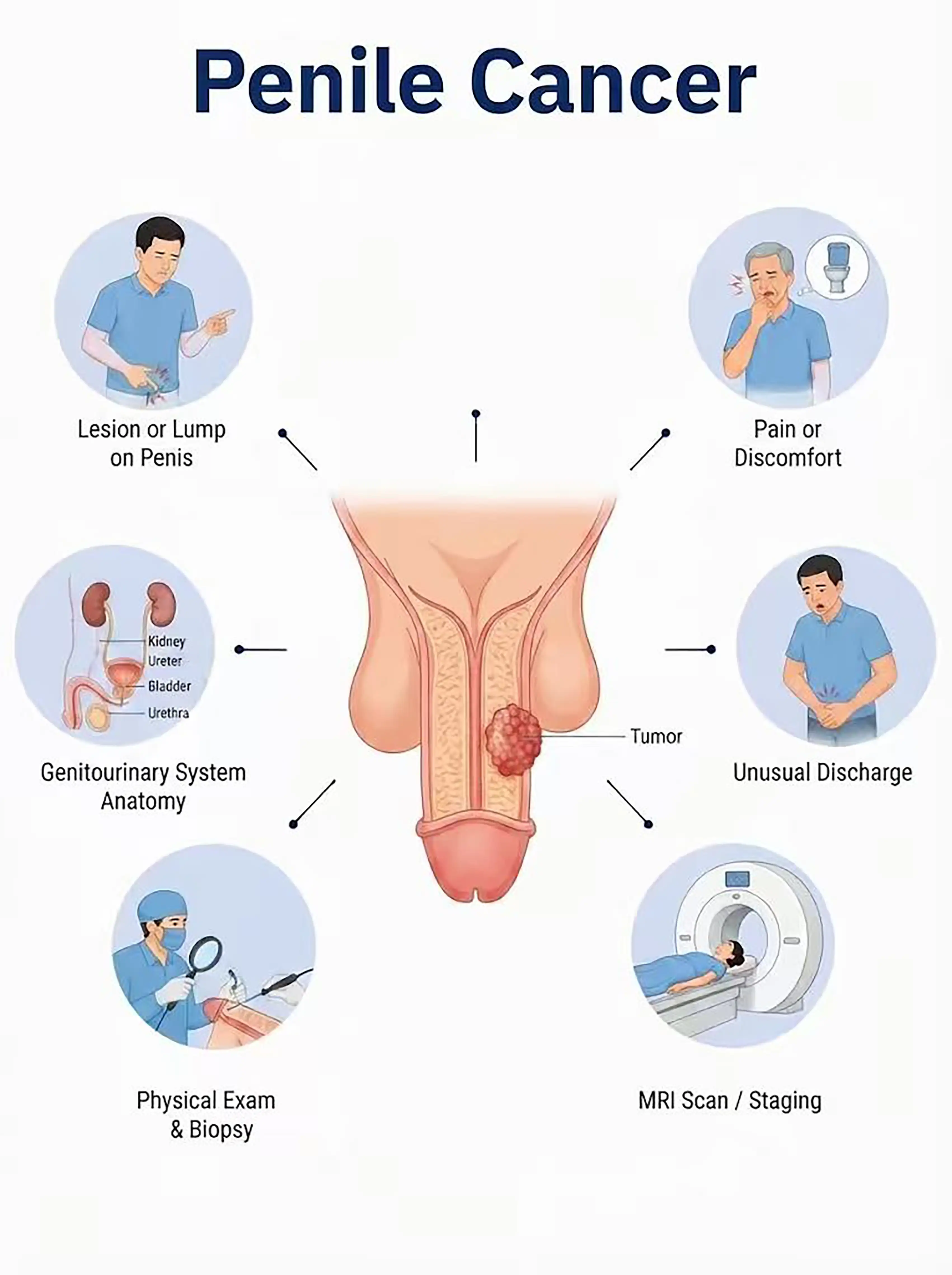
Penile cancer: staging and diagnosis
1Early diagnosis of penile cancer
Erythema, papules, ulcers, or white patches appear on the glans penis or foreskin. Clinical examination reveals suspicious lesions;an incisional biopsy is performed to establish a definitive pathological diagnosis.
2.Diagnosis of intermediate-stage penile cancer
The lesion enlarges to form a cauliflower-like mass or a deep ulcer.Biopsy is used to determine the pathological type and degree of differentiation, while ultrasound assesses invasion of the corpus cavernosum and inguinal lymph node metastasis.
3.Diagnosis of advanced penile cancer
Tumor extensively invades the penile shaft and urethra, or metastasizes distantly to inguinal lymph nodes and the lungs.Whole-body CT and PET-CT assess the extent of metastasis.
If not diagnosed promptly, the tumor can extensively invade the penile shaft and carries a high risk of lymph node metastasis; advanced stages may require total penectomy, severely affecting quality of life.
When to Seek Medical Attention
Early signs of penile cancer include red patches, ulcers, lumps, and abnormal discharge on the glans penis.Please consult a doctor via the "Chat with a Doctor" feature for early detection.During the online consultation, the doctor will ask about your symptoms and the condition of your foreskin, and guide you regarding a biopsy.
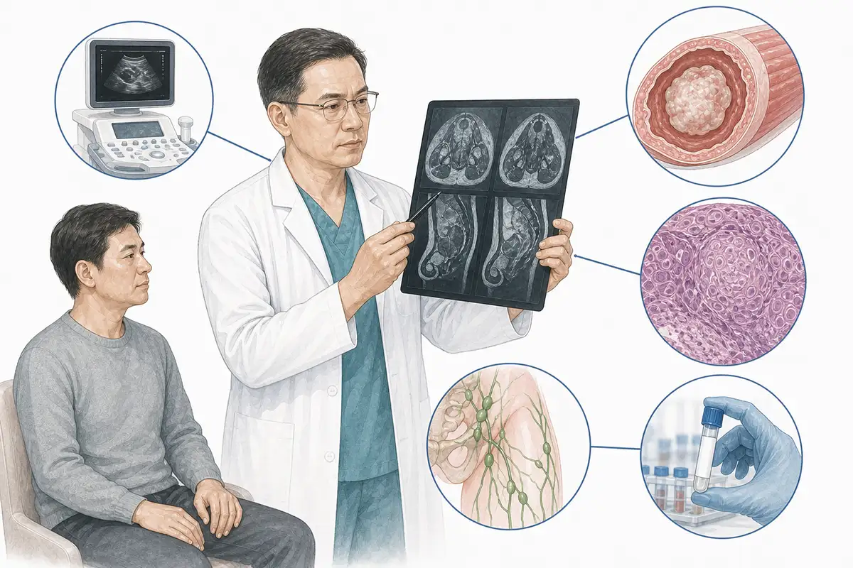
Basis for diagnosing penile cancer
1.Clinical examination
Comprehensive visual inspection and palpation of the glans penis, foreskin, and coronal sulcus—evaluating the morphology, size, location, and mobility of lesions—form the basis for initial diagnosis.
2.Histopathological biopsy
Biopsy of suspicious tissue for pathological examination is the gold standard for diagnosing penile cancer; it identifies the pathological type (squamous cell carcinoma accounts for over 95%) and the degree of differentiation.
3.Penile ultrasound
High-frequency ultrasound assesses tumor depth of invasion, involvement of the corpus cavernosum, and the status of surrounding tissues, serving as an important tool for local staging.
4.HPV test
Test for HPV infection status (specifically HPV types 16 and 18); approximately 40%–50% of penile cancers are associated with HPV infection, and this test aids in etiological analysis.
5.Inguinal lymph node assessment
Palpation and ultrasound are used to assess for inguinal lymph node enlargement, guide fine-needle aspiration biopsy of suspicious nodes, and evaluate metastatic status.
6.Pelvic MRI
MRI assesses the relationship of the tumor with the corpus cavernosum, urethra, and surrounding tissues, holding significant value for the staging of locally advanced tumors.
7.Chest and abdominal CT
Assesses for distant metastases in the lungs, liver, and retroperitoneal lymph nodes; a standard examination for initial staging.
8.Whole-body PET-CT
Assess for distant metastasis; offers advantages in detecting occult metastatic lesions that are difficult to identify via conventional imaging.
9.Sentinel lymph node biopsy
Inject a tracer to locate and biopsy the sentinel lymph node; this assesses the status of lymph node metastasis in early-stage tumors and guides the extent of surgery.
Daily Self-Check
Monitor the glans penis for erythema, ulcers, masses, or abnormal discharge. Maintain local hygiene;individuals with a long foreskin should pay special attention to cleaning the area.
Conclusion
Penile cancer is diagnosed through clinical examination and tissue biopsy.Early detection allows for sexual function-sparing surgery and a favorable prognosis.


