


Vulvar cancer is a tumor formed by the malignant proliferation of squamous epithelial cells in the vulvar skin or mucosa.The vulva—comprising the mons pubis, labia majora, labia minora, clitoris, and vaginal vestibule—is the external portion of the female reproductive organs.
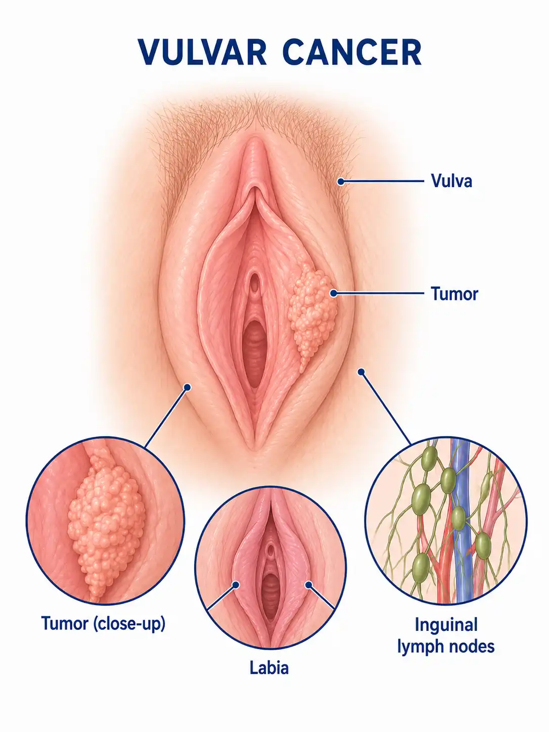
Global overview of vulvar cancer
Vulvar cancer accounts for approximately 4%–5% of malignancies in the female reproductive system; it is most common in elderly women, with a peak incidence between the ages of 65 and 75. In recent years, the incidence among younger women has risen, a trend associated with HPV infection.
Consult a doctor regarding cancer-related issues
Women experiencing symptoms such as vulvar itching, skin whitening, vulvar lumps, or non-healing ulcers should promptly consult a gynecologic oncologist.
Main types of vulvar cancer
1. Squamous cell carcinoma
It is the most common type of vulvar cancer, accounting for approximately 85%–90% of cases; subtypes include keratinizing, non-keratinizing, and basaloid types.
2. Melanoma
Originates in vulvar melanocytes; accounts for approximately 2%–5% of vulvar cancers. Highly malignant with a poor prognosis; commonly occurs on the labia minora and clitoris.
3. Basal cell carcinoma
Relatively rare; slow-growing with a low metastasis rate and good prognosis; most commonly found on the labia majora.
4. Paget's disease
A type of intraepithelial adenocarcinoma characterized by eczema-like changes; it may be associated with deep-seated invasive adenocarcinoma.
5. Adenocarcinoma subtype
Originates in vulvar glands, such as Bartholin's glands or sweat glands; relatively rare. Includes adenocarcinoma and adenoid cystic carcinoma.
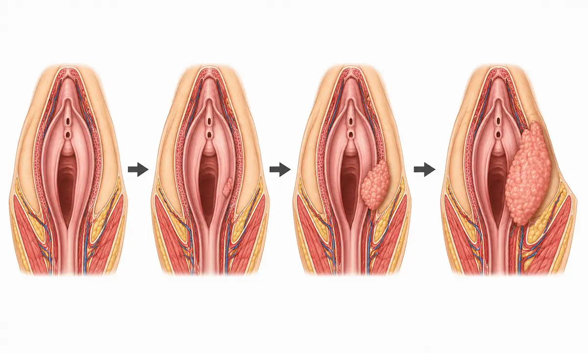
Typical symptoms and signs of vulvar cancer
1. Vulvar itching
Persistent vulvar itching is the most common symptom; it is chronic and resistant to treatment, requiring differentiation from vulvar dystrophy.
2. Skin whitening
The appearance of white plaques or areas of skin thickening and whitening warrants vigilance for vulvar intraepithelial neoplasia or early-stage invasive carcinoma.
3. Vulvar mass
Detection of a nodule or mass in the vulvar region; the lesion is firm in texture, and the surface may appear cauliflower-like or ulcerated.
4.Non-healing ulcer
A persistent ulcer or area of erosion with irregular margins that bleeds easily upon contact raises a high suspicion of malignancy.
5. Pain and bleeding
In the advanced stage, persistent pain and contact bleeding occur; invasion of the urethra or rectum by the tumor causes corresponding symptoms.
6. Inguinal mass
Enlarged, hard inguinal lymph nodes with poor mobility suggest possible lymph node metastasis.
What are the diagnostic methods for vulvar cancer
1. Gynecological examination
Carefully inspect and palpate all areas of the vulva, observing skin color and checking for masses, ulcers, or white lesions to assess the extent of the lesion.
2. Pathological biopsy
Wedge resection biopsy of suspicious lesions is the gold standard for diagnosing vulvar cancer and must be performed under local anesthesia.
3. Colposcopy
Colposcopy allows for magnified observation of vulvar lesions and guides biopsy sites, helping to detect early, minute lesions.
4. Pelvic MRI
MRI allows for the assessment of tumor invasion depth, the relationship with the urethra and anus, and the status of inguinal lymph node metastasis.
5. HPV testing
Test for high-risk HPV infection status; squamous cell carcinoma in young patients is frequently associated with HPV infection.
6. Sentinel lymph node biopsy
In early-stage patients, sentinel lymph node biopsy can assess lymph node metastasis status, avoiding unnecessary lymph node dissection.
Treatment Methods for Vulvar Cancer
1. Wide local excision
Early-stage tumors may be treated with wide local excision, removing the tumor along with 1–2 cm of surrounding normal tissue, while preserving the appearance and function of the vulva as much as possible.
2. Radical vulvectomy
For large tumors or multiple lesions, radical vulvectomy is required, potentially combined with partial resection of the urethra or rectum if necessary.
3. Inguinal lymph node dissection
For patients with suspected lymph node metastasis, inguinal-femoral lymph node dissection may be performed either synchronously or in stages.
4. Radiotherapy
Postoperative adjuvant radiotherapy is indicated for patients with lymph node metastasis or positive surgical margins to reduce the rate of local recurrence.
5. Chemotherapy
Platinum-based chemotherapy can be used for the systemic treatment of patients with advanced or recurrent disease.
6. Concurrent chemoradiotherapy
Concurrent chemoradiotherapy is an important treatment option for patients with locally advanced, unresectable disease.
7. Targeted therapy
The use of drugs targeting specific molecular markers for advanced vulvar cancer is currently being explored.
8. Follow-up monitoring
Postoperative follow-up involving regular vulvar examinations and inguinal palpation is crucial to monitor for local recurrence and lymph node metastasis.
Conclusion
Vulvar cancer is a common malignant tumor of the female reproductive system, associated with HPV infection and vulvar dystrophy.With early detection, standardized surgery, and necessary adjuvant therapy, the prognosis is favorable.


