


Vulvar cancer accounts for 3%–5% of malignancies in the female reproductive tract and is relatively rare.It predominantly affects postmenopausal women over the age of 65. Approximately 50% of cases are associated with HPV infection, while the other 50% are linked to chronic vulvar dystrophy (such as lichen sclerosus).
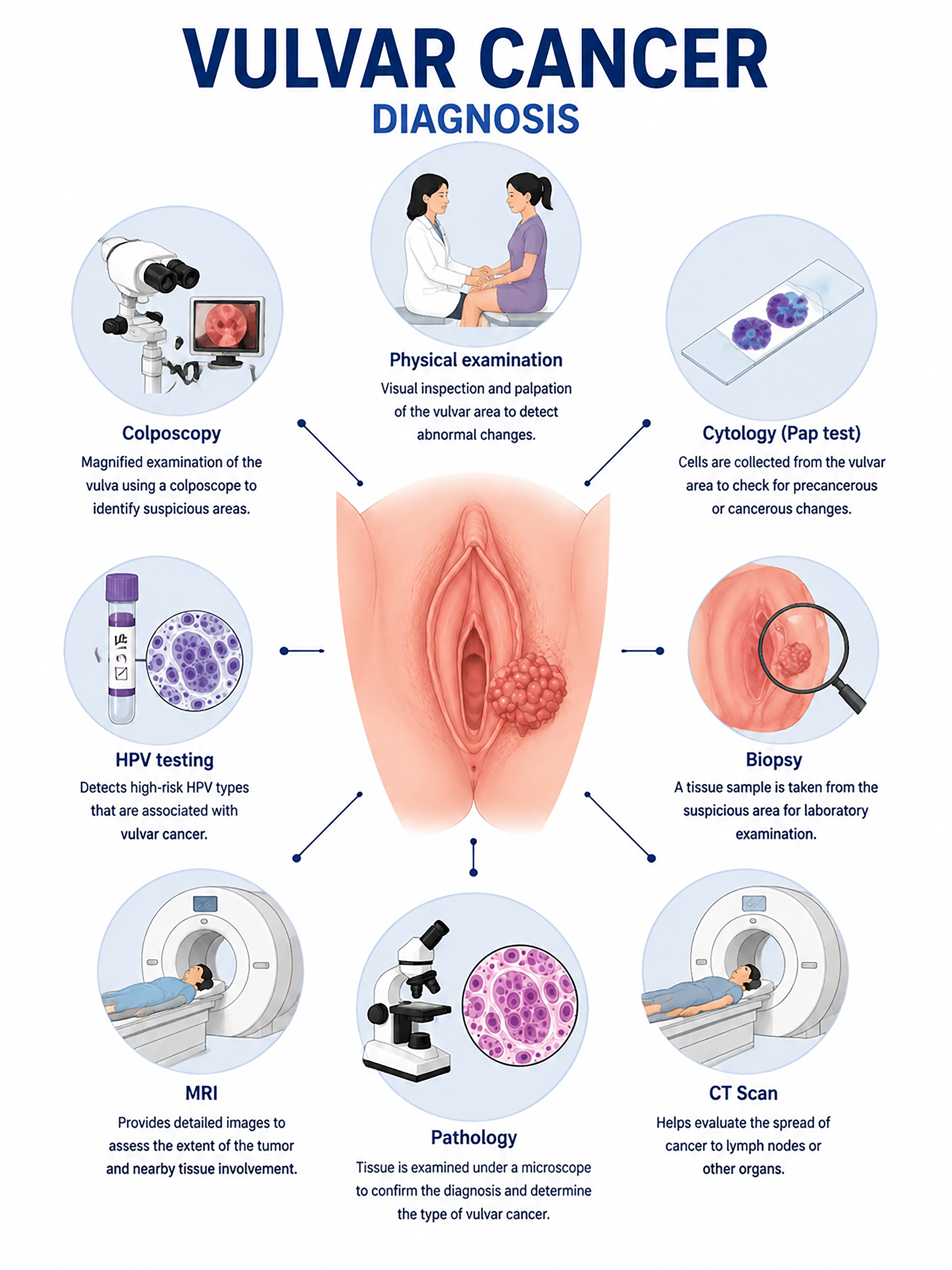
Diagnosis of vulvar cancer stages
1.Early diagnosis of vulvar cancer
The tumor is confined to the vulva (<2 cm), presenting with vulvar itching, leukoplakia (white patches), or ulceration.Lesions are observed via colposcopy, and the pathological diagnosis is confirmed by biopsy.
2.Diagnosis of intermediate-stage vulvar cancer
Enlargement of a vulvar lump or ulcer.Perform multiple biopsies to determine the pathological type and degree of differentiation; use MRI to assess the depth of tumor invasion and involvement of surrounding tissues.
3.Diagnosis of advanced vulvar cancer
Tumor invasion of the urethra or vagina, or distant metastasis to inguinal lymph nodes and the lungs.Whole-body CT and PET-CT are used to assess the extent of metastasis.
If vulvar cancer is not diagnosed promptly, the tumor may invade the urethra and vagina; there is a high rate of metastasis to inguinal lymph nodes, and the prognosis worsens significantly.
When to Seek Medical Attention
Early signs of vulvar cancer include itching of the vulva, changes in skin color, ulcers, and lumps.Please consult a doctor via "Chat with a Doctor" for early detection.During the online consultation, the doctor will ask about your symptoms and their duration, and guide you through the process of a gynecological examination and biopsy.
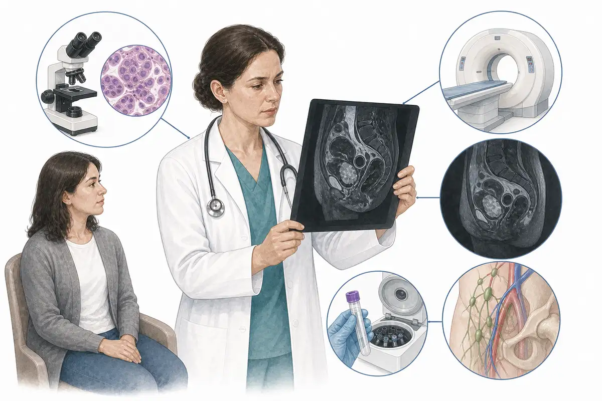
Basis for diagnosing vulvar cancer
1.Histopathological biopsy
Biopsy of suspicious tissue for pathological examination is the gold standard for diagnosing vulvar cancer; it identifies the pathological type (squamous cell carcinoma, melanoma, etc.) and the degree of differentiation.
2.Colposcopy
Specialized magnification is used to examine the vulvar mucosa; acetic acid and iodine tests are employed to localize suspicious lesions and guide precise biopsies.
3Pelvic MRI
MRI evaluates tumor size, depth of infiltration, and the relationship with the urethra and vagina, serving as an important imaging tool for local staging.
4.Contrast-enhanced CT scan
Contrast-enhanced CT to evaluate inguinal and pelvic lymph node metastasis, as well as the presence of distant organ metastasis.
5.HPV testing
Test for high-risk HPV infection status; approximately 50% of vulvar cancers are associated with HPV infection, aiding in etiological analysis and prognostic assessment.
6.Sentinel lymph node biopsy
Inject a tracer to locate and excise the sentinel lymph node; this assesses lymph node metastasis status and allows patients with early-stage disease to avoid unnecessary inguinal lymph node dissection.
7.Colposcopy
Assess for synchronous or metachronous intraepithelial neoplasia or carcinoma in the vagina and cervix; patients with vulvar cancer often present with concurrent HPV-related lesions in other sites.
8.Cystoscopy
For tumors located near the urethra, assessment of urethral invasion is conducted to guide the formulation of the surgical plan.
9.Whole-body PET-CT
Assess for distant metastasis; offers advantages in detecting occult metastatic lesions that are difficult to identify via conventional imaging, aiding in precise staging.
Daily Self-Check
Seek medical attention promptly if you experience persistent vulvar itching, white skin patches, ulcers, or lumps.Regularly self-examine the vulva for changes and consult a doctor if any abnormalities are noted.
Conclusion
Diagnosis of vulvar cancer relies on tissue biopsy and imaging-based staging.Early detection allows for a good prognosis through localized surgery, whereas advanced stages require comprehensive treatment.


