


Acoustic neuroma is a tumor formed by the abnormal proliferation of vestibular schwannoma cells.The vestibular nerve is part of the eighth cranial nerve and is responsible for transmitting auditory and balance information.
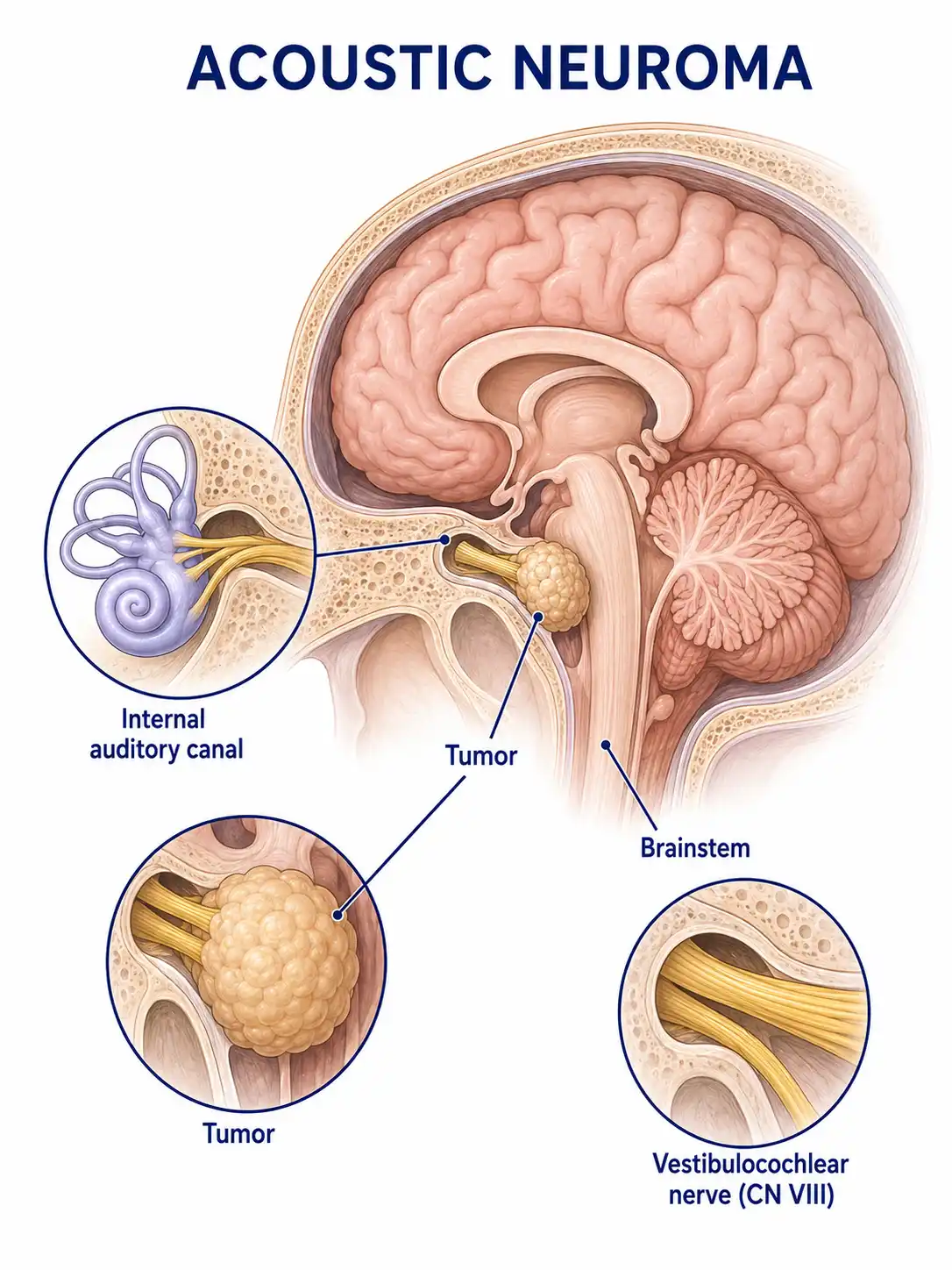
Global overview of acoustic neuroma
Acoustic neuromas account for approximately 8% of intracranial tumors and are the most common tumors in the cerebellopontine angle.They are usually unilateral and sporadic, whereas bilateral acoustic neuromas are associated with neurofibromatosis type 2.
Consult a doctor regarding cancer-related issues
Individuals experiencing symptoms such as progressive unilateral hearing loss, persistent tinnitus, vertigo, or facial numbness should promptly consult an ENT specialist or a neurosurgeon.
Main types of acoustic neuroma
1. Sporadic acoustic neuroma
The vast majority of cases are unilateral and lack a familial genetic background; onset typically occurs between the ages of 40 and 60, with variable growth rates.
2. Neurofibromatosis type 2
Bilateral acoustic neuromas are a characteristic manifestation of NF2; associated with NF2 gene mutations; typically presents at an early age.
3. Solid acoustic neuroma
The tumor is predominantly solid and firm in texture; surgery is relatively difficult due to tight adhesions to surrounding nerves.
4. Cystic acoustic neuroma
The tumor contains cystic components and may grow rapidly; the accumulation of cystic fluid can lead to a rapid worsening of symptoms.
5. Mixed-type acoustic neuroma
It contains both solid and cystic components; its clinical presentation and surgical difficulty fall somewhere between the two types.
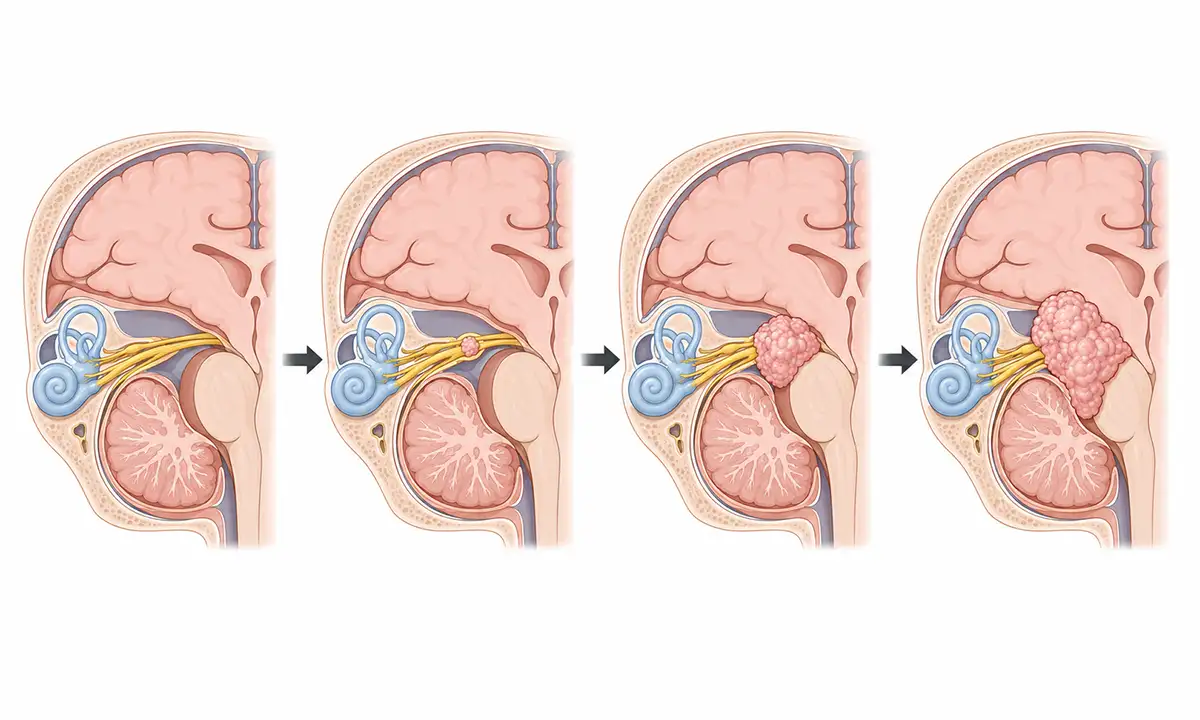
Typical symptoms and signs of acoustic neuroma
1. Hearing loss
Gradual unilateral sensorineural hearing loss is the most common initial symptom; it often goes unnoticed in the early stages but severely impairs communication in later stages.
2. Tinnitus
Persistent, high-pitched tinnitus—resembling the sound of cicadas—occurs in the ear on the affected side and serves as an early warning sign of an acoustic neuroma.
3. Vertigo
Tumor compression of the vestibular nerve causes balance disorders, vertigo, and a sense of instability, usually of mild severity.
4. Facial numbness
Tumor enlargement compressing the trigeminal nerve causes facial sensory loss, numbness, and pain; reduced corneal sensation may lead to exposure keratitis.
5. Facial paralysis
Compression of the facial nerve by the tumor results in facial muscle weakness, incomplete eyelid closure, and drooping of the corner of the mouth, severely affecting facial appearance.
6. Symptoms involving the lower cranial nerves
Compression of the lower cranial nerves by a giant tumor results in hoarseness, dysphagia, and choking when drinking.
Diagnostic methods for acoustic neuromas
1. Hearing test
Pure-tone audiometry reveals sensorineural hearing loss and reduced speech recognition on the affected side, serving as a crucial initial screening tool.
2. Auditory brainstem evoked potentials
ABR testing showing prolonged wave I-III latency or absent waveforms on the affected side is a sensitive method for screening for acoustic neuroma.
3. Internal auditory canal MRI
Contrast-enhanced MRI is the gold standard for diagnosing acoustic neuroma and can clearly detect small tumors (less than 5 mm) within the internal auditory canal.
4. Cranial CT
CT can demonstrate enlargement of the internal auditory canal and bone resorption, allowing for the assessment of the relationship between the tumor and surrounding bony structures.
5. Facial nerve function assessment
Assess facial nerve function grading; provides baseline data for surgical planning and the evaluation of postoperative functional recovery.
6. Vestibular function testing
Assesses vestibular function status, providing a basis for compensatory training following the complete loss of vestibular function on the affected side.
Treatment methods for acoustic neuroma
1. Observation and follow-up
For small or asymptomatic acoustic neuromas, or in elderly patients, periodic MRI monitoring may be chosen to track the tumor's growth rate.
2. Surgical treatment
Microsurgical tumor resection is the primary treatment modality, involving approaches such as the translabyrinthine, middle cranial fossa, and retrosigmoid approaches.
3. Stereotactic radiotherapy
Stereotactic radiotherapy, such as Gamma Knife, is suitable for small tumors or postoperative residual tumors; it can control tumor growth while preserving hearing.
4. Hearing preservation technique
Intraoperative neurophysiological monitoring and meticulous microsurgical techniques help preserve residual hearing function.
5. Facial nerve repair
Nerve anastomosis or grafting is performed when the facial nerve is damaged, aiming to restore facial expression function as much as possible.
6. Vestibular rehabilitation
Postoperative vestibular rehabilitation training helps patients adapt to their balance status following the loss of unilateral vestibular function.
7. Auditory aids
For patients with total hearing loss, auditory assistive devices such as cochlear implants or bone-anchored hearing aids may be considered.
8. Follow-up monitoring
Postoperative or post-radiotherapy follow-up involving regular MRI is crucial to monitor for residual tumor or recurrence.
Conclusion
Acoustic neuroma is the most common benign tumor in the cerebellopontine angle, primarily presenting with unilateral hearing loss and tinnitus.Microsurgery or stereotactic radiotherapy can control the tumor while maximizing the preservation of nerve function.


