


Acoustic neuromas account for 6%–8% of intracranial tumors and are the most common tumors in the cerebellopontine angle. They are predominantly unilateral and sporadic, whereas bilateral cases are associated with neurofibromatosis type 2 (NF2).They typically occur in individuals aged 40–60 and grow slowly, with an annual growth rate of approximately 1–2 mm.
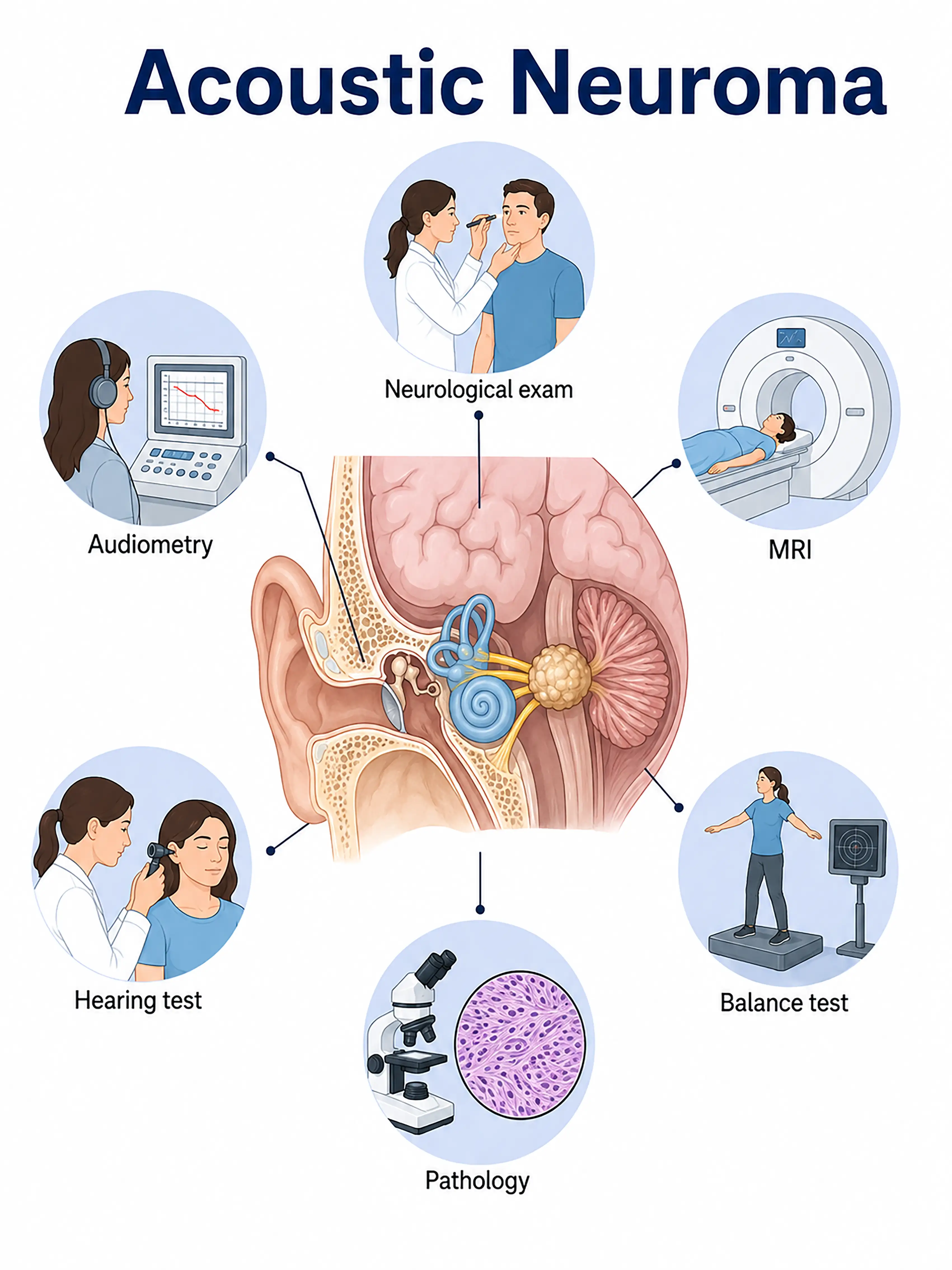
Staging-based diagnosis of acoustic neuroma
1.Early diagnosis of acoustic neuroma
Unilateral hearing loss and tinnitus are the initial symptoms.Contrast-enhanced MRI reveals a small nodule (<1.5 cm) within the internal auditory canal or cerebellopontine angle, showing marked, homogeneous enhancement and an "ice-cream cone" shape.
2.Diagnosis of intermediate-stage acoustic neuroma
Hearing declines further, accompanied by balance disorders and facial numbness.MRI shows tumor enlargement (1.5–3 cm), compression of the brainstem and cerebellum, and widening of the cisterns.
3.Diagnosis of advanced acoustic neuroma
A giant tumor causing severe brainstem compression, hydrocephalus, and cranial nerve damage.MRI reveals a large space-occupying lesion (>3 cm) with significant mass effect and ventricular enlargement.
If not diagnosed promptly, large acoustic neuromas can compress the brainstem, leading to life-threatening hydrocephalus, while compression of the facial nerve can result in permanent facial paralysis.
When to Seek Medical Attention
Early signs of acoustic neuroma include unilateral hearing loss, persistent tinnitus, and balance disorders.Please consult a doctor via the "Chat with a Doctor" feature to facilitate early detection.During the online consultation, the doctor will ask about changes in your hearing and your symptoms, and guide you through the process of undergoing hearing tests and an MRI.
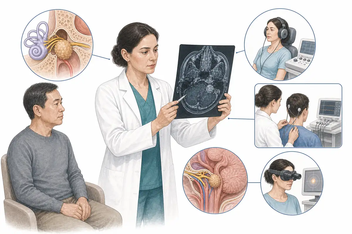
Diagnostic criteria for acoustic neuromas
1.Contrast-enhanced MRI of the inner ear
Contrast-enhanced MRI is the gold standard for diagnosing acoustic neuroma.Post-contrast T1-weighted imaging (T1WI) reveals a markedly enhancing mass within the internal auditory canal or cerebellopontine angle, often described as having an "ice-cream cone" shape, and clearly shows the relationship between the tumor, the facial nerve, and the brainstem.
2.Pure-tone audiometry
Standard audiological testing reveals unilateral sensorineural hearing loss, with speech recognition scores disproportionately lower than pure-tone thresholds; this is a characteristic audiological feature of acoustic neuroma.
3.Auditory brainstem response
ABR testing showing prolonged wave I-V latency or absent waveforms on the affected side is of reference value for screening for small, early-stage acoustic neuromas.
4.Vestibular function test
ENG/VNG examinations assess vestibular function; reduced or absent vestibular function on the affected side helps determine the impact of the tumor on the vestibular nerve.
5.Facial nerve function assessment
The House-Brackmann grading system is used to assess facial nerve function; recording baseline function preoperatively provides a reference for postoperative comparison.
6.CT bone window scan
CT reveals enlargement of the internal auditory canal and bone resorption; assessing the size of the canal provides a reference for selecting the surgical approach.
7.Multimodal MRI assessment
DTI reveals the spatial relationship between the facial nerve and the tumor, while fMRI assesses auditory cortex function, aiding in surgical planning and the preservation of function.
8.Brainstem auditory evoked potentials
Intraoperative monitoring of brainstem auditory evoked potentials (BAEP) allows for real-time assessment of brainstem function, thereby enhancing surgical safety.
9.Assessment of tumor growth rate
Serial MRI follow-ups are used to assess the annual tumor growth rate; active intervention is recommended for tumors growing at >3 mm/year, whereas those growing at <2 mm/year may be managed via observation and follow-up.
Daily Self-Check
Seek medical attention promptly if unilateral hearing loss or tinnitus occurs, especially if symptoms progressively worsen.Avoid mistaking unilateral hearing loss for age-related hearing loss or otitis media.
Conclusion
Diagnosis of acoustic neuromas relies on contrast-enhanced MRI and audiological examinations.Early detection helps preserve hearing and facial nerve function, thereby improving quality of life.


