


Soft tissue sarcomas are a group of rare malignant tumors comprising over 50 subtypes, accounting for 1% of all adult malignancies.They occur most frequently in the extremities (60%), followed by the retroperitoneum (15%–20%).Liposarcoma and leiomyosarcoma are the most common subtypes.Tumors with a high FNCLCC grade are associated with a poorer prognosis.
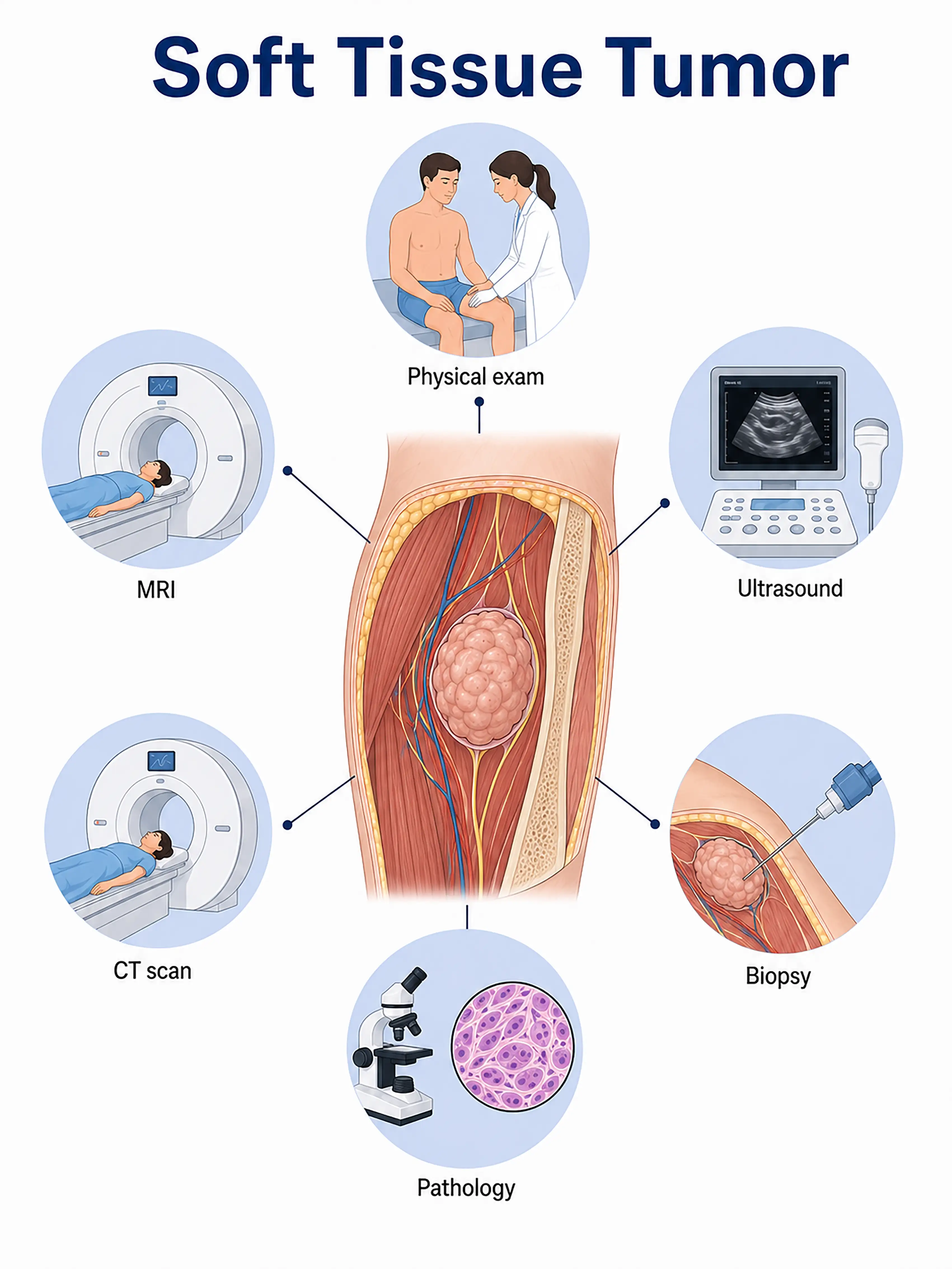
Staging and diagnosis of soft tissue tumors
1.Early diagnosis of soft tissue tumors
Small tumors (<5 cm) typically present as painless masses.MRI reveals the tumor's location, size, and relationship with surrounding tissues, while ultrasound-guided needle biopsy determines the pathological type and grade.
2.Diagnosis of intermediate-stage soft tissue tumors
The mass enlarges (>5 cm) or causes pain.MRI is used to assess tumor extent and involvement of vital structures; CT scans evaluate for lung metastasis; and biopsy confirms the histological subtype and grade.
3.Diagnosis of advanced soft tissue tumor
Extensive tumor invasion or distant metastasis to the lungs and liver.Whole-body CT and PET-CT assess the extent of metastasis; surgical acquisition of a complete specimen clarifies the pathology.
If soft tissue tumors are not diagnosed in time, high-grade sarcomas can progress rapidly and metastasize to distant sites, resulting in the loss of surgical resection opportunities and a significantly poorer prognosis.
When to Seek Medical Attention
Early signs of soft tissue tumors include the discovery of a painless lump, gradual enlargement of the lump, and localized pain.Please consult a doctor via "Chat with a Doctor" for early detection.During the online consultation, the doctor will ask about changes in the lump and guide you through MRI and biopsy procedures.
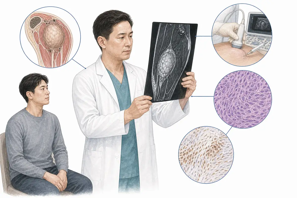
Basis for diagnosing soft tissue tumors
1.MRI examination
MRI is the preferred imaging modality for soft tissue sarcoma; it reveals the tumor's location, size, and boundaries, as well as its relationship with surrounding muscles, blood vessels, and nerves, serving as a core tool for surgical planning.
2.Needle biopsy
Obtaining tumor tissue samples for pathological examination via ultrasound- or CT-guided needle biopsy is the gold standard for diagnosing soft tissue sarcoma, allowing for the determination of histological subtype and FNCLCC grade.
3.CT scan
Contrast-enhanced CT evaluates tumor blood supply, bone invasion, and pulmonary metastasis; chest CT is a routine examination to rule out pulmonary metastasis.
4.Ultrasound examination
Ultrasound evaluates mass size, location, cystic-solid composition, and blood flow signals, guides needle biopsy positioning, and serves as a convenient tool for initial screening.
5.Pathological grading
Assessing the degree of tumor differentiation, mitotic count, and necrosis proportion using the FNCLCC system (Grades I–III) serves as a crucial basis for prognostic evaluation and treatment decision-making.
6.Chest CT scan
Assesses for pulmonary metastases; high-grade soft tissue sarcomas are prone to pulmonary metastasis, making this a standard examination for initial staging and follow-up.
7.PET-CT assessment
Whole-body PET-CT assesses distant metastasis, local recurrence, and chemotherapy response; high-grade sarcomas exhibit high metabolic activity.
8.Immunohistochemistry
Immunohistochemical analysis is performed on pathological specimens to differentiate between histological subtypes and guide precision therapy.
9.Molecular genetic testing
Detection of characteristic chromosomal translocations and fusion genes (e.g., FUS-CHOP, TLS-CHOP) to assist in pathological classification and the selection of targeted therapies.
Daily Self-Check
Seek medical attention promptly if a painless mass exceeding 5 cm in diameter and showing continuous growth is found anywhere on the body.Avoid massaging or applying heat to the mass yourself.
Conclusion
The diagnosis of soft tissue sarcoma relies on MRI and needle biopsy.Prompt medical attention should be sought to determine the nature of any unexplained mass, and early surgery is crucial.


