


Pancreatic cancer is the 12th most common cancer worldwide but ranks 7th in mortality.Approximately 66,000 new cases are diagnosed annually in the United States.The five-year survival rate is only about 12%, and approximately 80% of patients have unresectable tumors at the time of diagnosis.Smoking, chronic pancreatitis, and diabetes are major risk factors.
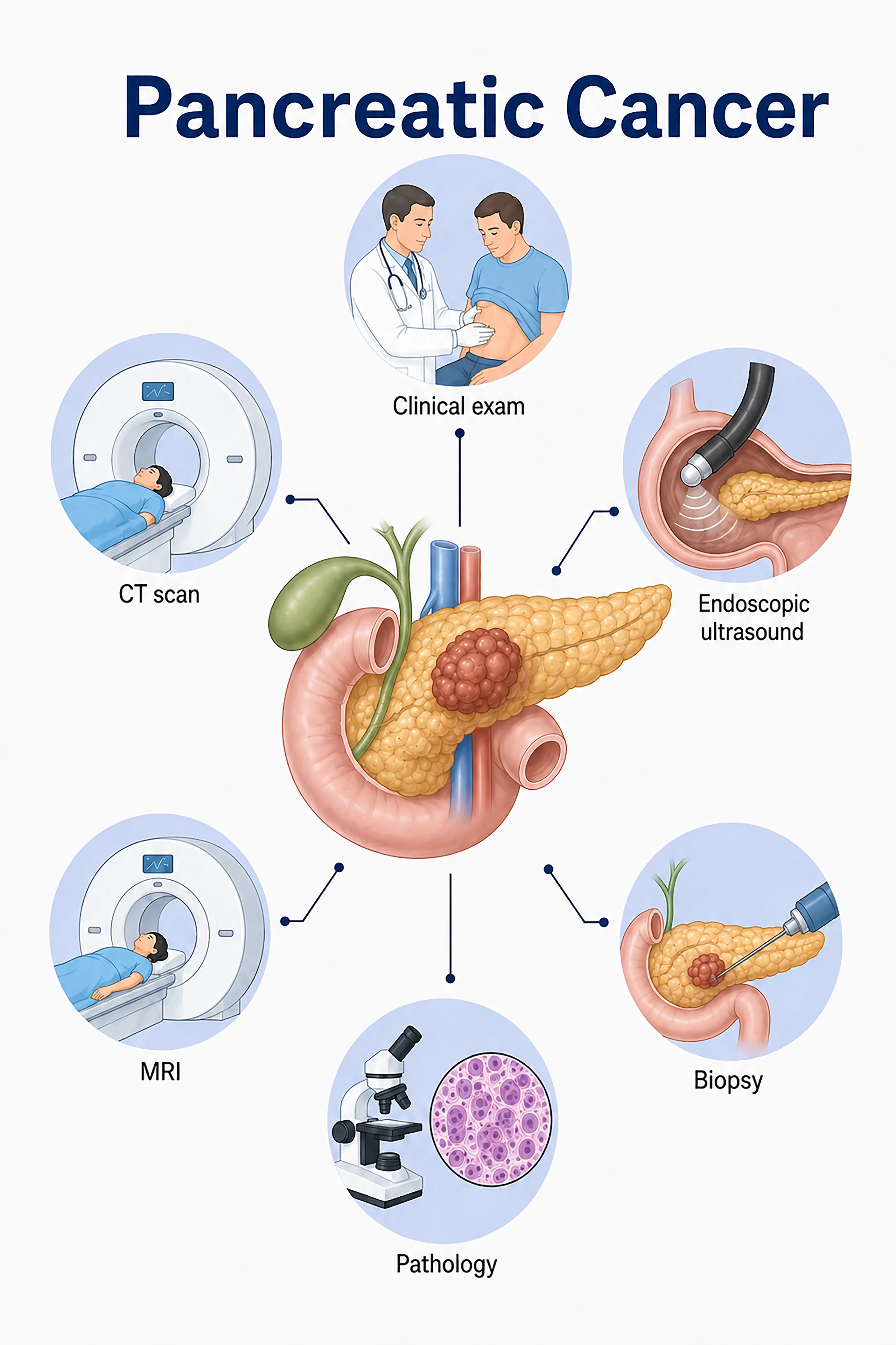
Diagnosis based on pancreatic cancer stages
1.Early diagnosis of pancreatic cancer
The tumor is confined to the pancreas (<2 cm) and is often asymptomatic.Contrast-enhanced CT reveals a hypodense lesion accompanied by pancreatic duct dilation; CA19-9 levels are mildly elevated; diagnosis is confirmed via EUS-guided needle biopsy.
2.Diagnosis of mid-stage pancreatic cancer
Presents with jaundice, abdominal pain, and weight loss.Contrast-enhanced CT and MRI reveal the extent of the tumor and vascular invasion, while EUS-FNA is used to obtain tissue samples for a definitive pathological diagnosis.
3.Diagnosis of advanced pancreatic cancer
Tumor invasion of major blood vessels or distant metastasis to the liver and peritoneum.Whole-body CT and PET-CT assess the extent of metastasis; multiple biopsies clarify the molecular subtype to guide chemotherapy regimens.
Without timely diagnosis, approximately 80% of patients are at an unresectable, advanced stage by the time of diagnosis; the five-year survival rate is extremely low, making it one of the malignancies with the poorest prognosis.
When to Seek Medical Attention
Early signs of pancreatic cancer include jaundice, upper abdominal pain, weight loss, and new-onset diabetes.Please consult a doctor via the "Chat with a Doctor" feature to facilitate early detection.During the online consultation, the doctor will ask about your symptoms and risk factors, and guide you through necessary tests such as CT scans and endoscopic ultrasound.
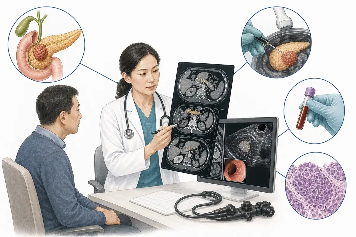
Basis for diagnosing pancreatic cancer
1.Contrast-enhanced CT scan
Contrast-enhanced CT is the preferred imaging modality for diagnosing pancreatic cancer, revealing low-density pancreatic masses, pancreatic duct dilation, and vascular invasion; it is a core tool for staging and assessing surgical resectability.
2.Endoscopic ultrasound examination
EUS allows for close-range observation of pancreatic lesions and ultrasound-guided fine-needle aspiration (FNA) for tissue sampling; it is the most reliable minimally invasive method for the definitive diagnosis of pancreatic cancer.
3.CA19-9 test
Carbohydrate antigen 19-9 (CA19-9) is the most important serum marker for pancreatic cancer; elevated levels suggest the possibility of pancreatic cancer, and the marker can be used to assist in diagnosis and monitor treatment efficacy.
4.Abdominal MRI
MRI excels in visualizing pancreatic lesions; MRCP clearly displays the pancreaticobiliary ductal system, allowing for the assessment of the degree of biliary obstruction and pancreatic duct dilation.
5.Percutaneous needle biopsy
CT-guided percutaneous needle biopsy of a pancreatic mass is indicated for patients unable to undergo EUS or for whom EUS has failed, in order to establish a pathological diagnosis.
6.ERCP examination
Endoscopic retrograde cholangiopancreatography (ERCP) can visualize pancreatic duct stenosis or interruption and allow for the collection of pancreatic juice for cytologic examination; it also enables stent placement to relieve biliary obstruction.
7.Chest CT scan
Assesses for metastases in the lungs and mediastinal lymph nodes; a standard examination for initial staging.
8.Whole-body PET-CT assessment
Assess for distant metastasis; offers advantages in detecting occult metastatic lesions that are difficult to identify via conventional CT, aiding in staging and treatment planning.
9.Liquid biopsy testing
Detects circulating tumor DNA (ctDNA) and circulating tumor cells (CTCs); as emerging tools for auxiliary diagnosis and monitoring treatment efficacy, they facilitate the non-invasive assessment of tumor burden.
Daily Self-Check
Seek medical attention promptly if unexplained jaundice, upper abdominal pain, weight loss, or new-onset diabetes occurs.High-risk individuals should undergo regular abdominal imaging screening.
Conclusion
Early diagnosis of pancreatic cancer is difficult; contrast-enhanced CT and EUS-FNA are key diagnostic tools.CA19-9 aids in diagnosis and monitoring, and early detection is the only way to improve survival rates.


