


Ovarian cancer has the highest mortality among female reproductive malignancies worldwide; approximately 70% of cases are advanced at diagnosis. Incidence in China is rising, with five-year survival around 40%. Early diagnosis is key to improving survival.
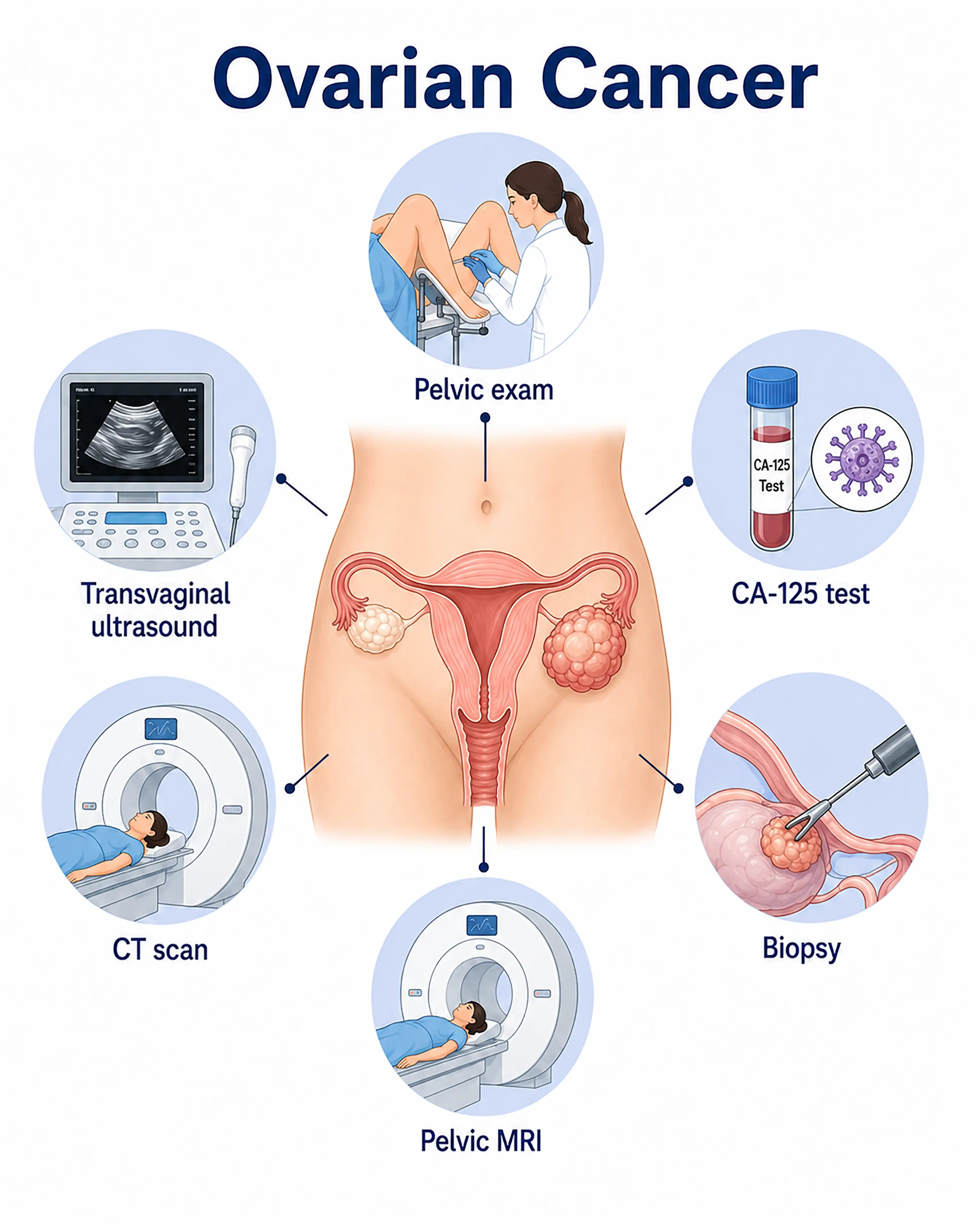
Ovarian cancer diagnosis by stage
1.Early-stage ovarian cancer diagnosis
Small tumors confined to the ovary usually cause no obvious symptoms. Pelvic ultrasound may incidentally identify a mixed cystic-solid adnexal mass, with mildly elevated CA125 and HE4, requiring close follow-up or surgical exploration.
2.Intermediate-stage ovarian cancer diagnosis
The tumor enlarges and ascites develops. Ultrasound and CT show a mixed cystic-solid pelvic mass with septa and papillary projections, and CA125 is markedly elevated. Laparoscopy can obtain tissue samples.
3.Advanced-stage ovarian cancer diagnosis
Extensive abdominal implantation and large-volume ascites develop. CT shows omental thickening, peritoneal nodules, and distant metastases. Exploratory laparotomy establishes stage and removes as much tumor as possible.
Without timely diagnosis, approximately 70% of ovarian cancers are advanced when detected, with extensive peritoneal implantation. The optimal surgical opportunity is missed and five-year survival decreases significantly.
When to Seek Medical Attention
Early warning signs include persistent bloating, an abdominal mass, indigestion, and unexplained weight loss. Please consult a doctor through "Chat with a Doctor" to support early detection. The doctor will ask about symptoms and family history and guide you through gynecological examination and ultrasound assessment.
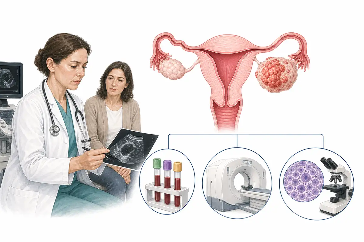
Basis for ovarian cancer diagnosis
1.Pelvic ultrasound
Transvaginal or abdominal ultrasound is the preferred screening method. It assesses ovarian size, shape, and mass characteristics, distinguishing cystic from solid lesions and identifying papillary projections and blood flow.
2.Tumor marker testing
Serum markers include CA125, HE4, and CEA. Elevated CA125 suggests possible epithelial ovarian cancer, and combining HE4 with CA125 improves diagnostic accuracy.
3.ROMA score assessment
The ROMA score combines CA125, HE4, and menopausal status to estimate malignancy risk in a pelvic mass and help determine whether surgical exploration is needed.
4.Pelvic CT and MRI
Contrast-enhanced CT and MRI clearly show tumor size, location, relationships with adjacent organs, and nodal metastases to assess surgical feasibility.
5.Exploratory laparoscopy
Laparoscopy directly examines pelvic and abdominal disease and obtains biopsies to establish pathology and stage and assess the feasibility of cytoreductive surgery.
6.Ascitic fluid cytology
Ascitic fluid is sampled for cancer cells. A positive result supports diagnosis and staging and is important evidence in advanced ovarian cancer.
7.Histopathological biopsy
Surgically removed or needle-sampled tumor tissue undergoes pathology, the gold standard for ovarian cancer diagnosis, identifying histological type and differentiation.
8.Whole-body PET-CT assessment
Assessing whole-body metastases detects small lesions missed by conventional imaging and guides treatment planning and prognosis assessment.
9.Genetic testing
Testing for susceptibility mutations such as BRCA1/2 helps assess disease risk and guide targeted therapy. Screening is recommended for patients with a family history.
Daily Self-Check
Monitor persistent bloating, abdominal distension, indigestion, and urinary frequency, especially in women with a family history of ovarian cancer. Regular gynecological examinations help detect pelvic abnormalities early.
Conclusion
Early ovarian cancer diagnosis is difficult, and approximately 70% of patients have advanced disease when diagnosed. Regular gynecological examinations and ultrasound screening support early detection. Unexplained bloating warrants prompt consultation.


