


Oral cancer refers to malignant tumors arising from the mucosa in various parts of the oral cavity, including cancers of the tongue, gums, buccal mucosa, floor of the mouth, hard palate, and lips; squamous cell carcinoma accounts for over 90% of these cases.Major risk factors include long-term smoking, alcohol consumption, betel nut chewing, HPV infection, poor oral hygiene, and chronic irritation.
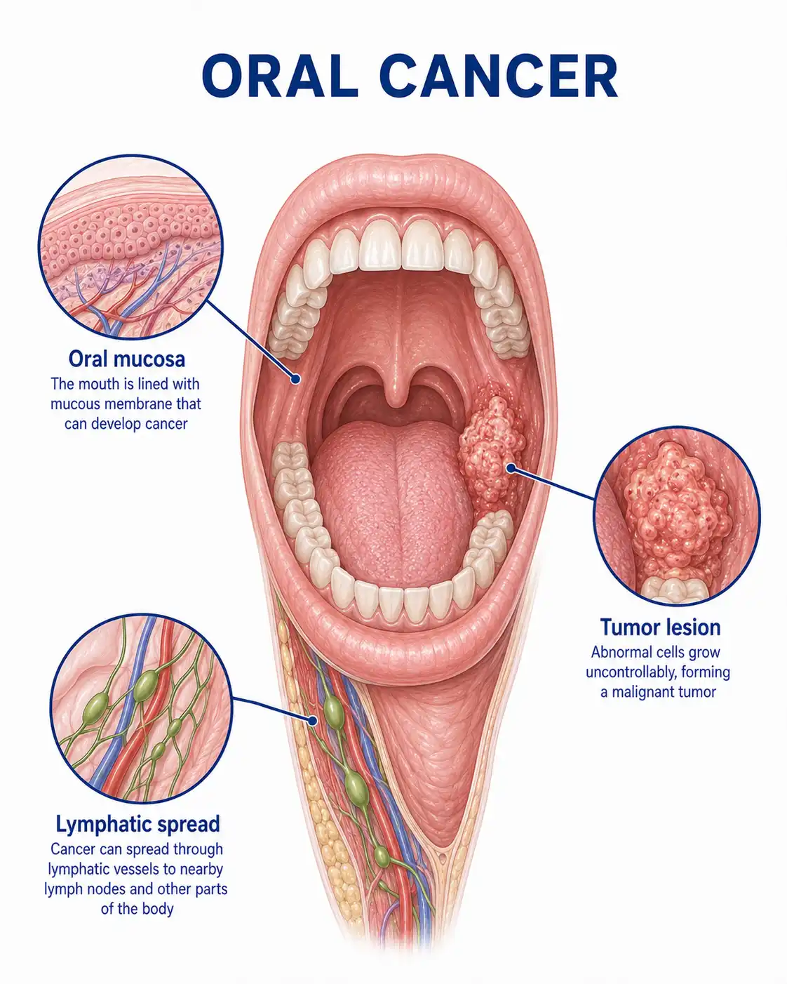
Global overview of oral cancer
Oral cancer is one of the most common malignant tumors worldwide, with over 350,000 new cases annually; incidence rates are higher in developing countries.In Southeast Asia, the incidence is significantly above the global average due to the habit of chewing betel nut.The incidence rate in men is approximately 2–3 times that of women; it most commonly affects individuals aged 40–60, though a trend toward occurrence in younger populations has been evident in recent years.
Consult a doctor regarding cancer-related issues
If an oral ulcer persisting for more than 2 weeks without healing, a white or red mucosal patch, unexplained pain, numbness, or a lump appears, one should promptly consult a department of oral and maxillofacial surgery or head and neck oncology.Following diagnosis, a multidisciplinary team should formulate an individualized treatment plan.
Main types of oral cancer
1.Tongue cancer
The most common type of oral cancer;it frequently occurs on the lateral borders and ventral surface of the tongue.Early signs include tongue ulcers or exophytic masses; it is highly invasive and prone to metastasis to cervical lymph nodes.
2.Gingival cancer
Occurs on the gingival mucosa of the upper or lower jaw, most commonly the lower gingiva.Early manifestations include gingival ulcers or papillary hyperplasia; it may invade the alveolar bone, causing tooth loosening and localized pain.
3.Buccal mucosa cancer
Occurs in the buccal mucosa; strongly associated with betel nut chewing and smoking.Often presents as a buccal ulcer or infiltrative mass, potentially invading the buccinator muscle and skin.
4.Carcinoma of the floor of the mouth
Occurs on the mucosa of the floor of the mouth; its concealed location makes early detection difficult.It tends to invade deep into the floor-of-mouth muscles and the jawbone, and often metastasizes to bilateral cervical lymph nodes.
5.Lip cancer
It most commonly affects the lower lip and is frequently associated with long-term sun exposure.It presents as an ulcer or a cauliflower-like mass at the vermilion border, grows relatively slowly, and has a favorable prognosis.
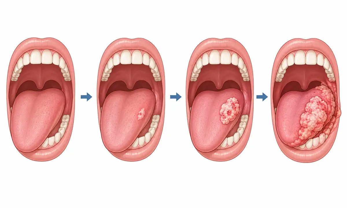
Typical symptoms and signs of oral cancer
1.Non-healing oral ulcer
An oral ulcer persisting for more than 2–3 weeks without healing; characterized by raised, crater-like edges, an irregular base, a tendency to bleed upon contact, and significant pain.
2.Oral leukoplakia
The appearance of white plaques or erythematous patches on the oral mucosa—characterized by a rough, thickened surface that cannot be wiped off—indicates a precancerous lesion or early-stage cancer; a biopsy is required for confirmation.
3.Oral mass or nodule
Hard masses or nodules appear in the oral cavity; the surface may resemble a cauliflower or have a papillary appearance. Pain and functional impairment may develop as the tumor grows.
4.Pain and numbness
Tumor invasion of nerves causes localized pain, a burning sensation, or numbness; tongue cancer may affect the lingual nerve, leading to referred pain in the ipsilateral ear and numbness of the tongue.
5.Dysphagia and speech disorders
Functional impairments such as dysphagia, slurred speech, and limited chewing ability occur when the tumor enlarges or invades the base of the tongue, the floor of the mouth, and pharyngeal structures.
6.Enlarged cervical lymph nodes
Oral cancer is prone to cervical lymph node metastasis, presenting as a painless mass in the neck (unilateral or bilateral) that is hard in texture and has poor mobility; this is a significant indicator of disease progression.
What are the diagnostic methods for oral cancer
1.Clinical oral examination
Visual inspection and palpation are used to examine changes in the color, texture, and morphology of the oral mucosa across various regions, thereby assessing the size, location, and extent of invasion of the primary tumor.
2.Tissue biopsy pathology
Biopsy or needle aspiration of suspicious tissue for pathological examination is the gold standard for diagnosing oral cancer; it identifies the pathological type and the degree of differentiation.
3.Neck ultrasound
High-frequency ultrasound evaluates the size, morphology, cortical thickening, and blood flow of cervical lymph nodes to determine the presence of metastasis and guide fine-needle aspiration biopsy.
4.Contrast-enhanced CT scan
Contrast-enhanced CT of the head and neck clearly delineates the extent of tumor invasion into surrounding soft tissues and the jawbone, and allows for the assessment of cervical lymph node and distant metastases.
5.Magnetic resonance imaging (MRI)
MRI offers high soft-tissue resolution, enabling precise assessment of the extent of tumor invasion into tongue muscles, the floor-of-mouth musculature, and neurovascular structures, thereby guiding the design of the surgical scope.
6.Systemic assessment/examination
Includes chest CT, abdominal ultrasound, and bone scanning to rule out distant metastases (e.g., to the lungs, liver, or bones), determine disease stage, and guide treatment planning.
Oral Cancer Treatment Methods
1.Surgical resection: Surgical resection is the primary treatment for oral cancer.Depending on tumor size and location, procedures such as wide local excision, combined jaw and neck radical resection, or partial jaw resection are selected, aiming to achieve radical resection.
2.Mohs micrographic surgery: Tumor tissue is excised layer by layer with real-time intraoperative pathological examination; this allows for precise control of the excision margin and maximal preservation of normal tissue, making it suitable for lip cancer and superficial lesions.
3.Radiotherapy: Monotherapy is suitable for early-stage patients unable to tolerate surgery, or it is used in combination with surgery for intermediate- to advanced-stage cases. Intensity-modulated radiotherapy (IMRT) allows for precise irradiation of the target area, reducing damage to surrounding normal tissues.
4.Chemotherapy: Neoadjuvant chemotherapy can shrink tumors to facilitate surgical resection, while adjuvant chemotherapy eliminates potential micrometastases.Commonly used drugs include cisplatin and 5-fluorouracil; chemotherapy is frequently administered concurrently with radiotherapy.
5.Targeted therapy: Molecular targeted drugs that act on targets such as the epidermal growth factor receptor (EGFR) can inhibit tumor growth and metastasis.Combining drugs like cetuximab with radiotherapy and chemotherapy can improve treatment outcomes for patients with advanced-stage disease.
6.Immunotherapy: Immune checkpoint inhibitors kill tumor cells by activating the body's immune system, demonstrating promising therapeutic efficacy and application prospects in advanced or recurrent/metastatic oral cancer.
7.Reconstructive surgery: Following extensive resection, free flap transplantation or local tissue flap repair is used to address the defect and restore oral contours as well as vital physiological functions such as chewing, swallowing, and speech.
8.Follow-up monitoring: Regular post-treatment re-examination of the oral cavity and neck to monitor for recurrence and metastasis.Quit smoking and drinking, maintain oral hygiene, avoid chewing betel nut, and undergo regular oral examinations for the early detection of second primary cancers.
Conclusion
Treatment for oral cancer primarily involves surgery, supplemented by comprehensive approaches such as radiotherapy, chemotherapy, targeted therapy, and immunotherapy.Early detection and treatment can significantly improve cure rates and quality of life; quitting smoking and alcohol, avoiding betel nut chewing, and undergoing regular oral check-ups are important preventive measures.


