


Oral cancer is the 16th most common cancer globally, accounting for 2%–3% of all cancer cases.In my country, the incidence of oral cancer is rising year by year.It most commonly affects men aged 40–60, with major risk factors including smoking, alcohol consumption, betel nut chewing, and HPV infection.The five-year survival rate can reach 80% for early-stage cases but falls below 30% for advanced-stage cases.
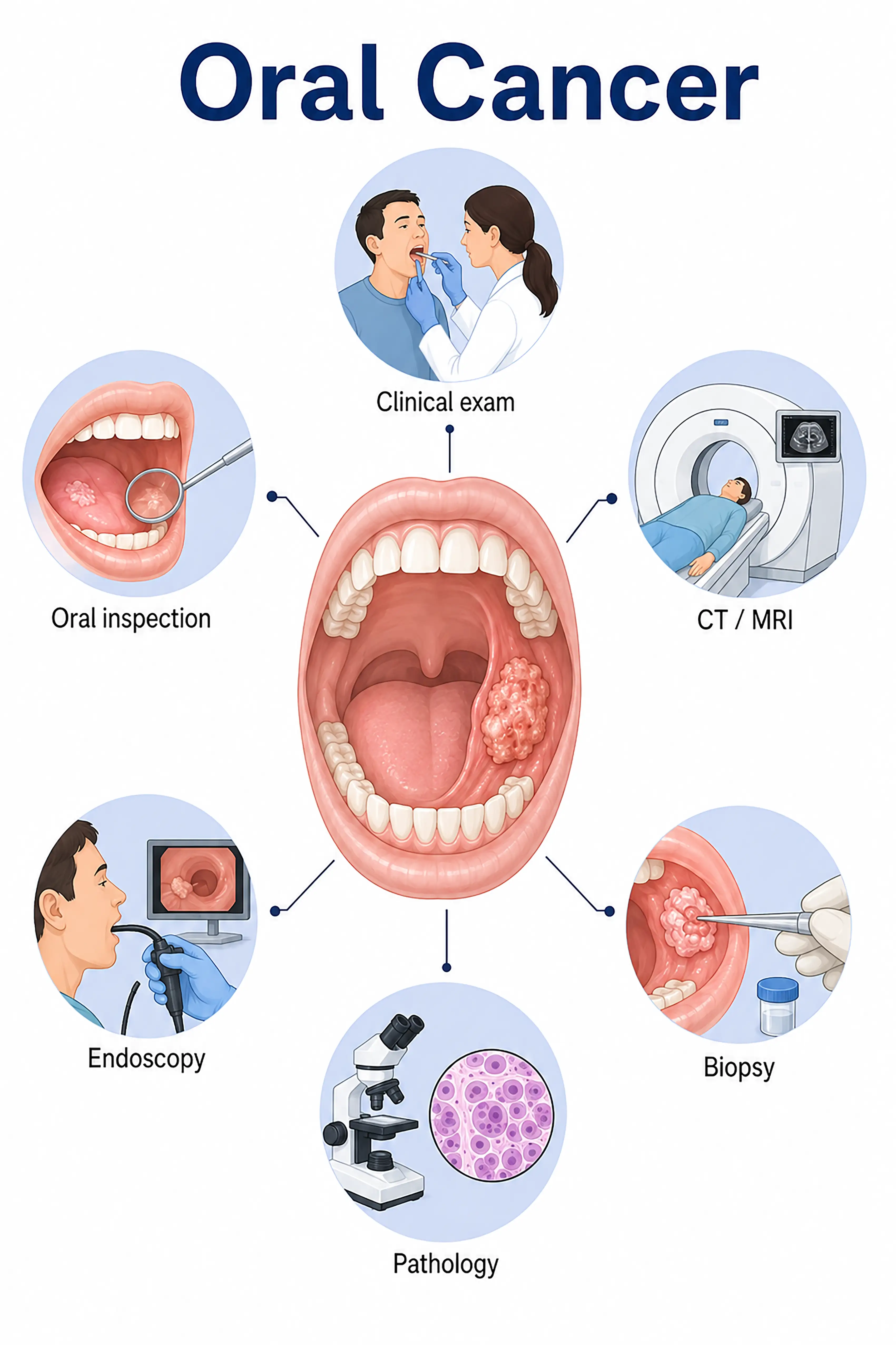
Diagnosis based on oral cancer stages
1.Early diagnosis of oral cancer
Presence of a persistent, non-healing oral ulcer, white patch, or red patch. Clinical visual inspection and palpation reveal mucosal abnormalities;diagnosis is confirmed via incisional biopsy and pathological examination.
2.Diagnosis of intermediate-stage oral cancer
The ulcer enlarges, accompanied by induration and pain.Biopsy is used to determine the pathological type and degree of differentiation, while contrast-enhanced CT or MRI assesses the depth of tumor invasion and cervical lymph node metastasis.
3.Diagnosis of advanced oral cancer
The tumor invades the jawbone or metastasizes to cervical lymph nodes.Whole-body CT and PET-CT are used to assess for distant metastasis, and multiple biopsies are performed to confirm the pathology.
If not diagnosed promptly, the tumor may invade the jawbone and masticatory muscles, causing difficulty in opening the mouth and eating; there is a high rate of metastasis to cervical lymph nodes, and the prognosis worsens significantly.
When to Seek Medical Attention
Early signs of oral cancer include mouth ulcers that do not heal for more than three weeks, white or red patches in the mouth, lumps, and pain.Please consult a doctor via "Chat with a Doctor" for early detection.During the online consultation, the doctor will ask about your oral hygiene habits and symptoms, and guide you through oral examinations and biopsies.
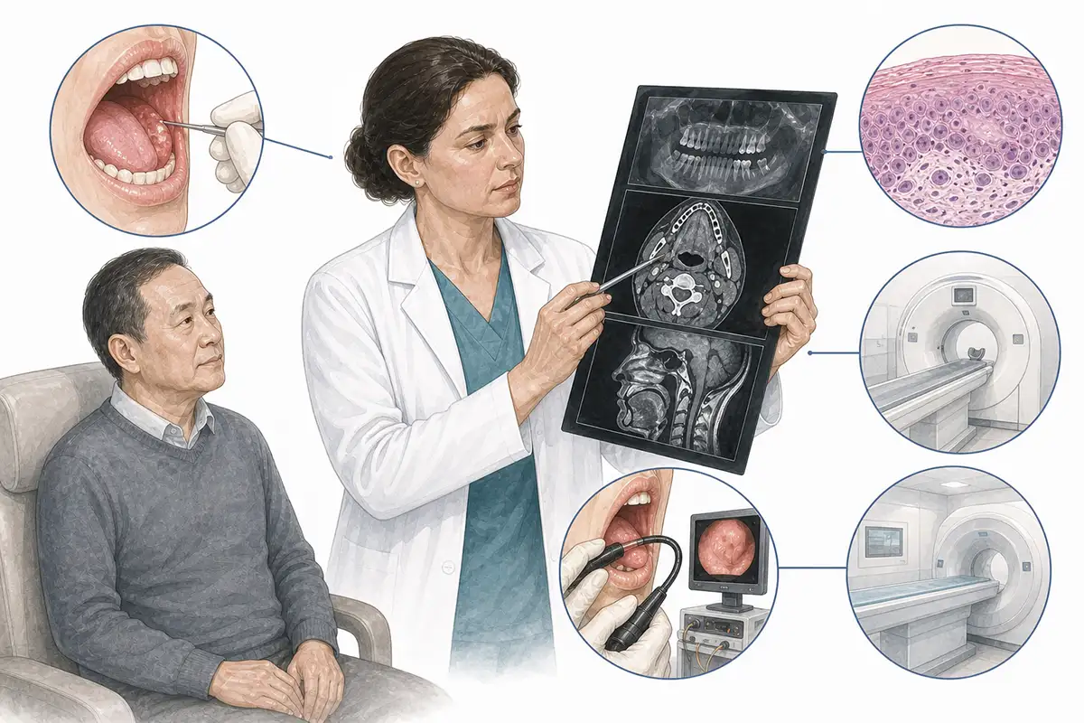
Basis for oral cancer diagnosis
1.Clinical oral examination
Visual inspection and palpation of various oral regions (tongue, buccal mucosa, gingiva, floor of the mouth, and hard palate)—observing for ulcers, masses, leukoplakia, or color changes—serve as the foundation for preliminary screening.
2.Histopathological biopsy
Biopsy of suspicious tissue for pathological examination is the gold standard for diagnosing oral cancer; it identifies the pathological type (95% are squamous cell carcinoma) and the degree of differentiation.
3.Contrast-enhanced CT scan
Contrast-enhanced CT evaluates tumor size, bone invasion, and cervical lymph node metastasis, serving as a core imaging tool for staging and surgical planning.
4.MRI examination
MRI offers high soft-tissue resolution, enabling the assessment of the tumor's relationship with tongue muscles, blood vessels, and nerves, as well as the differentiation between tumor tissue and inflammatory edema.
5.Cervical lymph node ultrasound
High-frequency ultrasound assesses cervical lymph node metastasis, guides biopsy of suspicious lymph nodes, and assists in staging and diagnosis.
6.Panoramic X-ray
Panoramic dental radiography assesses the mandible and maxilla for bone invasion and serves as a useful reference for determining whether a tumor has infiltrated the bone.
7.Whole-body PET-CT assessment
Assess for distant metastasis; offers advantages in detecting occult metastatic lesions that are difficult to identify via conventional imaging, aiding in precise staging.
8.HPV testing
Some oropharyngeal cancers are associated with HPV infection; HPV testing aids in determining the etiology, assessing prognosis, and making treatment decisions.
9.Endoscopy
Oral endoscopy aids in visualizing lesions in hidden areas, helping to detect early-stage abnormalities and guide precise biopsies.
Daily Self-Check
Monitor for persistent oral ulcers, leukoplakia, or masses.Avoid smoking, alcohol consumption, and betel nut chewing, and undergo regular oral examinations.
Conclusion
Oral cancer is diagnosed through clinical examination and pathological biopsy.Early detection allows for surgical cure, whereas the prognosis for advanced-stage disease is poor.


