


Global disease prevalence: Meningioma is one of the most common primary brain tumors worldwide; it is more frequently found in women, and most cases are benign and slow-growing.
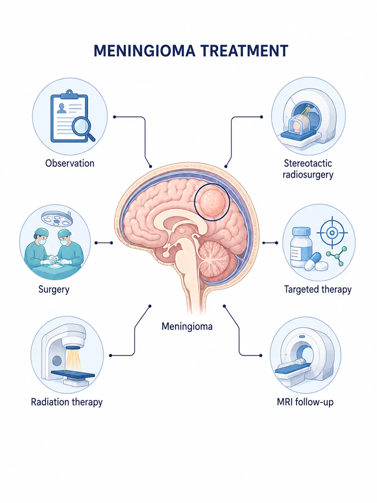
Staged treatment for meningioma
Early treatment (involving a localized, small tumor with a favorable response)
specifically surgical resection combined with adjuvant therapy—can effectively control tumor progression, and the patient's prognosis is relatively good.
Advanced-stage treatment (addressing severe compression, alleviating symptoms, and prolonging survival)
primarily employs systemic therapies such as radiotherapy to relieve symptoms of neurological compression and maximize patient survival time.
Consequences of delayed early treatment: Failure to receive timely, standardized treatment in the early stages allows the tumor to continue enlarging and compressing surrounding brain tissue and nerves; missing the optimal window for intervention leads to a worsening of the condition.
Why choose Fosun Health for meningioma treatment
Leveraging the CyberKnife M6, VitalBeam, neurosurgery, and MRI imaging platforms available at its affiliated hospitals, Fosun Health facilitates multidisciplinary consultations involving experts in neurosurgery, radiotherapy, medical imaging, pathology, and rehabilitation.Based on tumor size, growth rate, WHO grading, and the tumor's relationship with the visual pathway, brainstem, venous sinuses, and cranial nerves, the team selects the appropriate approach: active surveillance, microsurgery, or precision radiotherapy.
For eligible small lesions, the M6 CyberKnife offers a non-invasive (non-craniotomy) local treatment option; for larger tumors or those adjacent to critical structures, fractionated precision radiotherapy can be used to minimize radiation exposure to normal brain tissue and cranial nerves, thereby reducing the risk of certain neurological adverse reactions.Post-treatment care may include epilepsy management, assessment of visual and auditory functions, neurological rehabilitation, and long-term imaging follow-up.

Meningioma treatment methods
1.M6 CyberKnife treatment
Suitable for meningiomas that are small, well-defined, continuously growing, or symptomatic but unsuitable for surgery, as well as small residual or recurrent lesions post-surgery.
Treatment advantages:
① Targeting small tumors: Suitable for select localized intracranial and skull base lesions.
② Protect normal brain tissue: Reduce radiation dose to the brainstem, visual pathways, and cranial nerves.
③ No craniotomy required: Provides an option for patients at high surgical risk.
④ Short treatment course: Eligible patients can undergo single-fraction or hypofractionated treatment.
2.VitalBeam precision radiotherapy
Suitable for meningiomas that are large in volume, adjacent to the visual pathway or brainstem, residual post-surgery, high-grade, or unsuitable for CyberKnife hypofractionated therapy.
Treatment advantages:
① Suitable for large target volumes: Covers the tumor, the surgical bed, and high-risk areas of dural attachment.
② Reduce single-fraction dose: Suitable for lesions immediately adjacent to the optic nerve and brainstem.
③ Protection of brain tissue: Reduce radiation exposure to normal brain tissue through dose shaping.
④ Supports postoperative treatment: Used for high-grade or incompletely resectable meningiomas.
3.Microsurgical meningioma resection
Suitable for surgical candidates presenting with mass-effect symptoms, continuous growth, or large tumor volume, or those requiring definitive pathological grading.
4.Active surveillance
Small, asymptomatic meningiomas with low imaging-based risk profiles may be monitored via periodic MRI scans following specialist assessment.
5.Proton beam therapy
Uses proton beams to precisely target tumor tissue while maximizing protection of surrounding normal brain tissue; suitable for patients with meningioma in challenging locations....and consolidate therapeutic efficacy.
6.Pharmacotherapy
Controls tumor growth using hormones or targeted drugs; provides a treatment option for patients with meningioma who are unsuitable for surgery or radiotherapy.
7. Interventional therapy:
Indicated for certain large, highly vascular meningiomas scheduled for surgical resection; preoperative embolization of feeding arteries may be performed to reduce the risk of intraoperative hemorrhage.
Post-treatment care
Maintain a regular daily routine and a balanced diet; avoid strenuous exercise; attend scheduled follow-up appointments and actively cooperate with subsequent treatment as advised.
Post-treatment examinations
Periodic cranial MRI and neurological function assessments are required after treatment to timely detect tumor recurrence or changes in the disease status.
Conclusion
Although often benign, meningioma poses a threat that cannot be ignored; successful treatment hinges on early detection, diagnosis, and intervention, as well as selecting an appropriate treatment plan and adhering to follow-up care.


