


Meningiomas account for approximately 30%–40% of all intracranial tumors.The incidence rate is higher in women than in men (approximately 2:1), and they most commonly occur in individuals aged 40 to 70.The vast majority are WHO Grade I benign tumors, while approximately 5%–10% are atypical or malignant (WHO Grades II–III).They grow slowly, and some may remain asymptomatic for long periods.
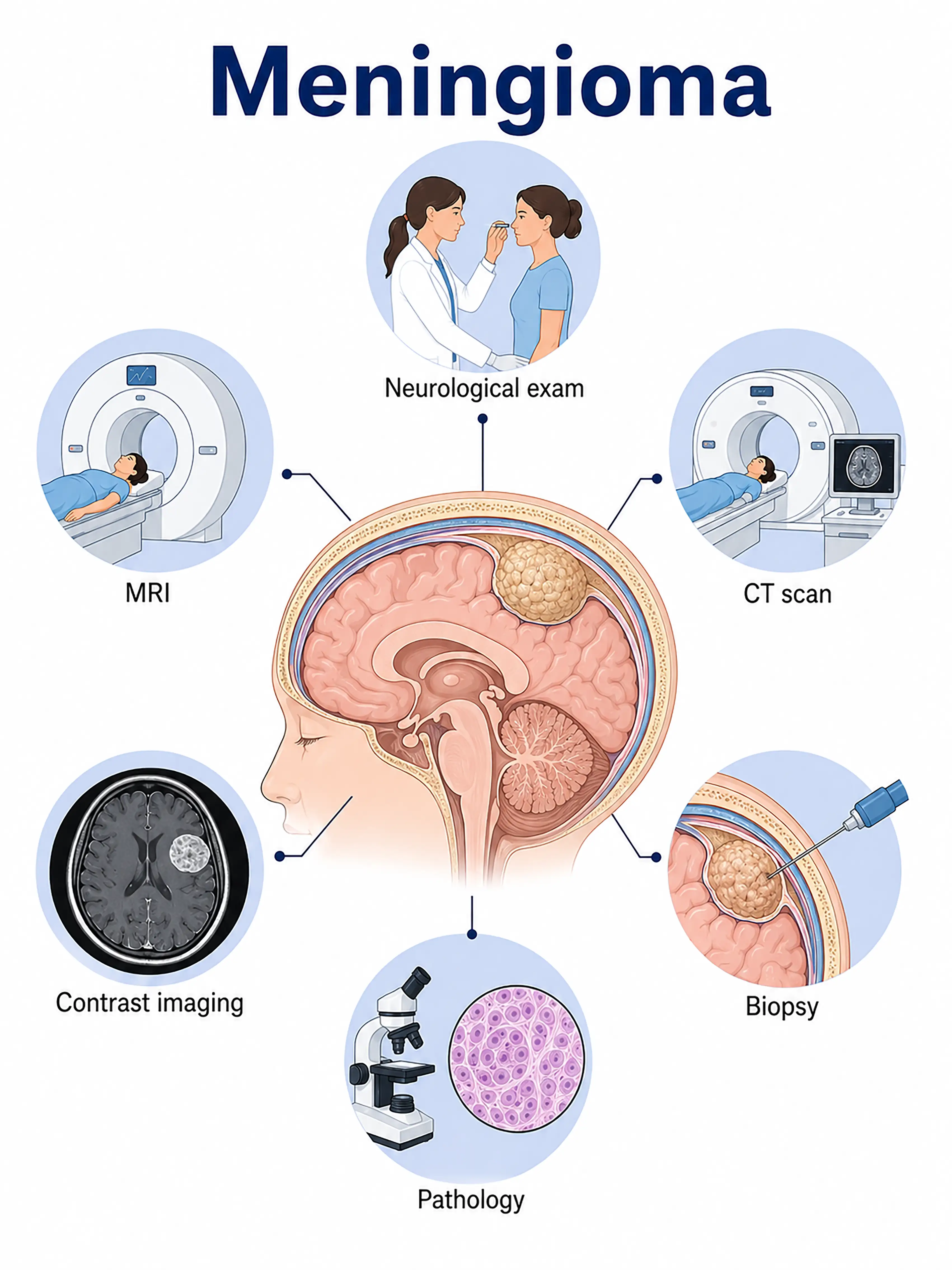
Diagnosis based on meningioma stages
1.Early diagnosis of meningioma
Small tumors, often without obvious symptoms.MRI shows a mass attached to the dura mater with isointense T1 signals and isointense or slightly hyperintense T2 signals; contrast-enhanced imaging shows marked, homogeneous enhancement and the "dural tail sign."
2.Diagnosis of intermediate-stage meningioma
Headache, seizures, or focal neurological deficits.MRI shows tumor enlargement with surrounding edema and mass effect;CT may reveal calcification and hyperostosis.
3.Diagnosis of advanced meningioma
A large tumor is causing severe intracranial pressure elevation.MRI shows a large mass with midline shift; surgical resection is required to determine the pathological grade (WHO Grade I–III).
If not diagnosed promptly, large meningiomas can compress vital brain regions, causing severe neurological impairment;malignant meningiomas can invade surrounding tissues and recur.
When to Seek Medical Attention
Early signs of meningioma include persistent headaches, seizures, vision changes, and limb weakness.Please consult a doctor via the "Chat with a Doctor" feature for early detection.During the online consultation, the doctor will ask about your symptoms and their progression, and guide you through the process of getting a cranial MRI.
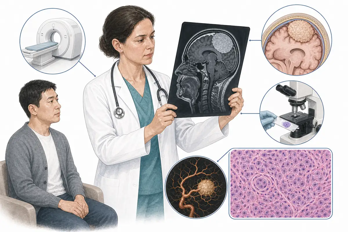
Basis for meningioma diagnosis
1.Cranial MRI (plain + contrast-enhanced)
MRI is the preferred method for diagnosing meningioma.Typical findings include an isointense mass attached to the dura mater, showing marked, homogeneous enhancement and the characteristic "dural tail sign."
2.Cranial CT scan
CT can demonstrate intratumoral calcification, hyperostosis, and changes in adjacent bone; it is superior to MRI for assessing bone invasion by skull base meningiomas and serves as an important adjunct to preoperative evaluation.
3.Histopathological examination
Pathological examination of the surgically resected specimen determines the WHO grade (Grades I–III) and molecular characteristics, guiding postoperative follow-up and adjuvant therapy regimens.
4.Cerebral angiography
For highly vascular meningiomas or tumors situated near major blood vessels, angiography is used to assess the tumor's blood supply and its relationship with critical vessels, aiding in surgical planning.
5.T1-weighted sagittal view
Sagittal T1-weighted contrast-enhanced scanning offers unique advantages in visualizing parasagittal, falcine, and sellar region meningiomas, allowing for the assessment of the tumor's relationship with the sagittal sinus.
6.PET-CT assessment
PET-CT can differentiate between low-grade and high-grade meningiomas and assess tumor metabolic activity, aiding in the distinction between tumor recurrence and radiation necrosis.
7.Visual acuity and visual field examination
For meningiomas in the sellar region or along the sphenoid ridge, comprehensive ophthalmic examinations—assessing visual acuity, visual fields, and fundus changes—are a crucial component of diagnosis and follow-up.
8.Neurological function assessment
A systematic neurological examination—assessing the tumor's impact on brain function, including motor, sensory, speech, and cranial nerve functions—forms the basis of the diagnostic assessment.
9.Molecular marker testing
Detection of NF2 gene mutations and alterations in genes such as TRAF7 and AKT1 assists in molecular classification and correlates with tumor biological behavior and prognosis.
Daily Self-Check
Seek medical attention promptly if unexplained headache, seizures, or vision changes occur.Postmenopausal women experiencing these symptoms should be alert to the possibility of a meningioma.
Conclusion
The vast majority of meningiomas are benign, and contrast-enhanced MRI is the key diagnostic tool.Complete surgical resection can achieve a cure for most patients.


