


Meningiomas are tumors resulting from the abnormal proliferation of meningeal cells.The meninges consist of three layers of connective tissue that encase and protect the brain and spinal cord; meningiomas most commonly originate from arachnoid granulation cells.
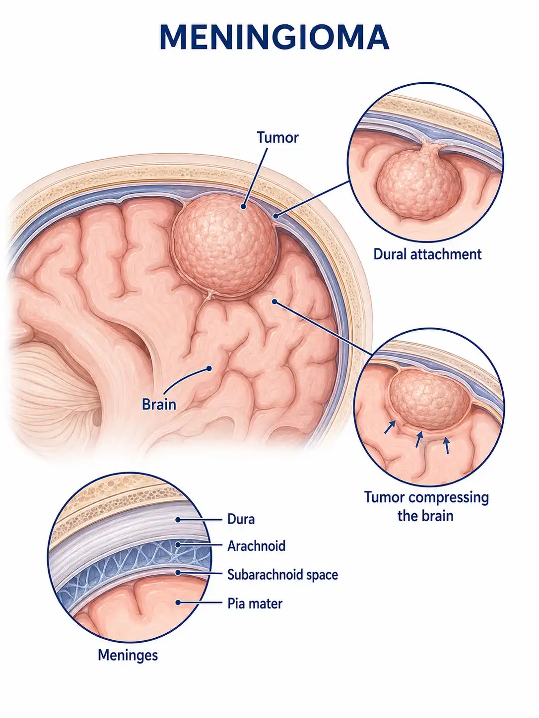
Global overview of meningioma
Meningioma has the highest incidence among all primary intracranial tumors;it is more common in women—particularly middle-aged and older women—with some cases linked to estrogen levels.
Consult a doctor regarding cancer-related issues
Individuals experiencing symptoms such as persistent headache, vision loss, seizures, or limb weakness, or those with an intracranial mass incidentally discovered during a physical examination, should promptly consult a neurosurgeon.
Main types of meningioma
1. Endothelial meningioma
WHO Grade I; the most common pathological subtype; cells are arranged in nests or whorls; mitotic figures are rare; slow-growing.
2. Fibrous meningioma
WHO Grade I; composed of spindle cells and abundant collagen fibers; firm in texture; slow-growing; prognosis is favorable.
3. Atypical meningioma
WHO Grade II; active cellular proliferation; increased mitotic figures; postoperative recurrence rate is higher than that of Grade I meningiomas.
4. Anaplastic meningioma
WHO Grade III;characterized by high malignancy, marked cellular atypia, a tendency for recurrence and metastasis, and a poor prognosis.
5. Multiple meningiomas
The simultaneous or sequential occurrence of two or more intracranial meningiomas may be associated with genetic factors or neurofibromatosis type 2.
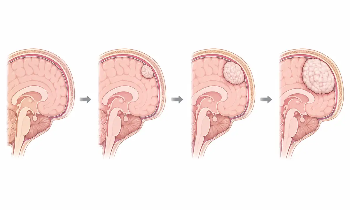
Typical symptoms and signs of meningioma
1. Headache
Tumor compression of brain tissue or elevated intracranial pressure causes headache, typically a chronic, dull pain that gradually worsens.
2. Vision loss
Tumor compression of the optic nerve or optic chiasm causes vision loss and visual field defects; severe cases can lead to blindness.
3. Epileptic seizures
Tumor irritation of the cerebral cortex can trigger epileptic seizures; this is one of the common initial symptoms of meningiomas.
4. Limb weakness
Meningiomas located near the motor cortex can cause weakness, numbness, or impaired mobility in the contralateral limbs.
5. Loss of sense of smell
Meningiomas located at the anterior skull base can compress the olfactory nerve, leading to a diminished or lost sense of smell.
6. Mental status changes
Frontal lobe meningiomas can cause psychiatric symptoms such as personality changes, inattention, and memory loss.
Diagnostic methods for meningioma
1. Neurological examination
Detailed examinations of visual acuity, visual fields, muscle strength, sensation, and reflexes are conducted to assess neurological deficits.
2. Cranial MRI scan
Contrast-enhanced MRI is the preferred method for diagnosing meningiomas; typical findings include a well-defined, homogeneously enhancing mass attached to the meninges.
3. Cranial CT scan
CT can reveal intratumoral calcification and hyperostosis, aiding in the evaluation of the relationship between the tumor and the skull.
4. Angiography
Cerebral angiography can assess the tumor's blood supply, providing a reference for formulating the surgical plan.
5. Pathological examination
Pathological examination following surgical tumor resection clarifies the pathological type and WHO grade, guiding subsequent treatment and follow-up.
6. Molecular testing
Detection of NF2 gene mutations and chromosomal abnormalities helps clarify genetic background and assess prognosis.
What are the treatment methods for meningioma
1. Surgical treatment
Surgical resection is the primary treatment for meningioma; Simpson Grade I resection can significantly reduce the recurrence rate.
2. Observation and follow-up
For asymptomatic small meningiomas, periodic MRI monitoring may be chosen, with follow-up scans performed annually to track changes in the tumor.
3. Stereotactic radiotherapy
Stereotactic radiotherapy, such as Gamma Knife, is suitable for small meningiomas or postoperative residual tumors; it can control tumor growth.
4. External beam radiotherapy
Postoperative adjuvant radiotherapy is indicated for atypical and anaplastic meningiomas to reduce the risk of recurrence.
5. Targeted therapy
Targeted therapies for relapsed/refractory meningioma, including anti-angiogenic agents, are currently under investigation.
6. Interventional embolization
Preoperative endovascular embolization reduces tumor blood supply, lowers the risk of intraoperative hemorrhage, and facilitates the surgical procedure.
7. Symptomatic and supportive care
Antiepileptic drugs are used to control seizures, while glucocorticoids reduce peritumoral edema and alleviate related symptoms.
8. Follow-up monitoring
Regular postoperative MRI follow-up is conducted to monitor for recurrence; low-grade tumors require annual scans, while high-grade tumors require more frequent follow-up.
Conclusion
Meningiomas are the most common primary intracranial tumors, and most are benign.With regular imaging follow-up and timely surgery or radiotherapy, most patients achieve a favorable long-term prognosis.


