


Lymphoma is a common hematologic malignancy, categorized into Hodgkin lymphoma and non-Hodgkin lymphoma.It affects all age groups, with a higher incidence in males than in females.Precision staging and treatment guided by PET-CT have resulted in a cure rate of over 80% for Hodgkin lymphoma.
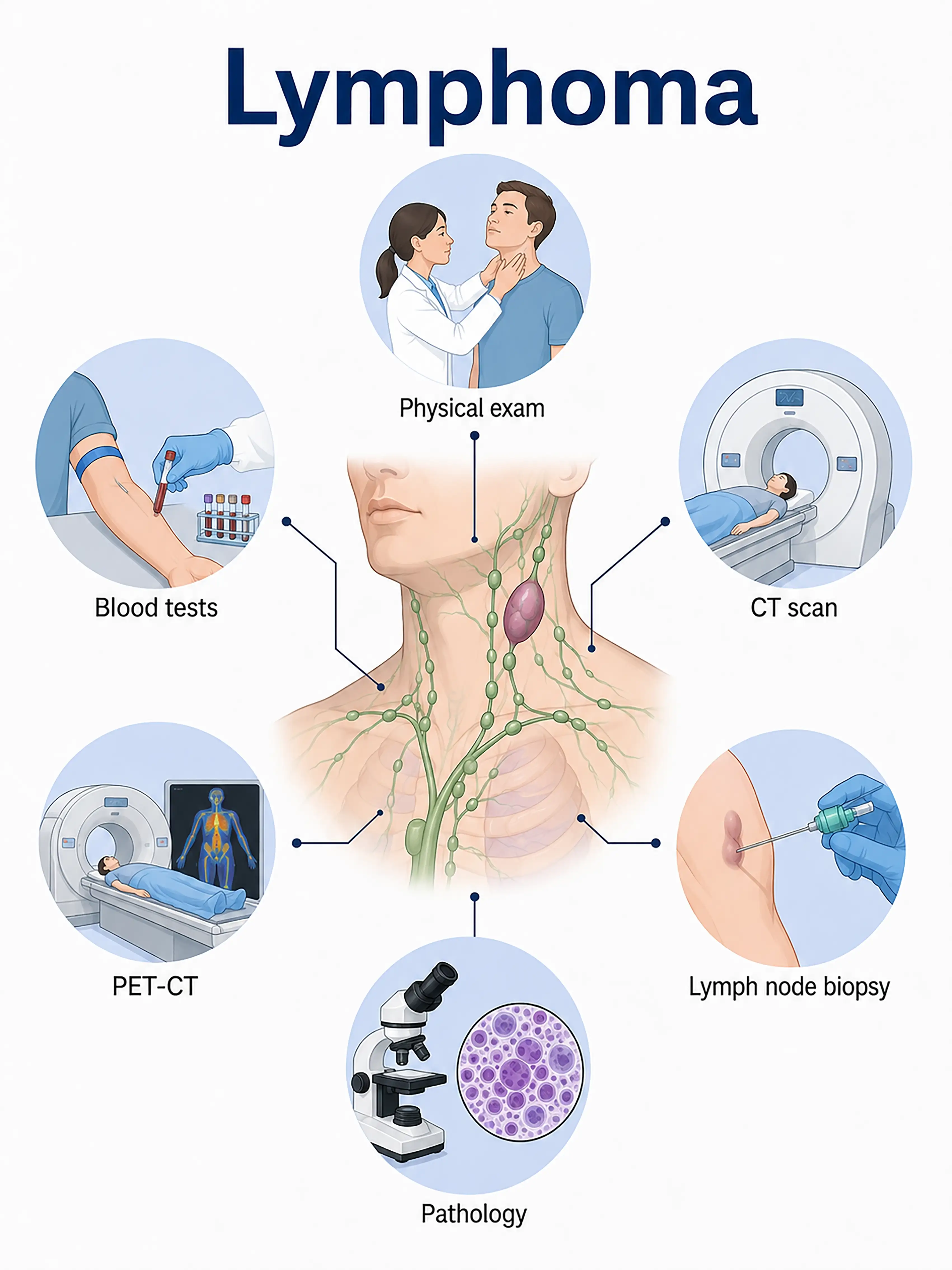
Lymphoma staging and diagnosis
1Early diagnosis of lymphoma
Painless lymphadenopathy (in the neck, axilla, or groin).Ultrasound is used to assess lymph node size and structure, while excisional biopsy of the intact lymph node is performed to determine the pathological type and immunohistochemical characteristics.
2.Intermediate-stage lymphoma diagnosis
Generalized lymphadenopathy accompanied by systemic symptoms (fever, night sweats, weight loss).CT and PET-CT scans are used to assess systemic lymph node and organ involvement, while bone marrow aspiration rules out bone marrow infiltration.
3.Advanced lymphoma diagnosis
Extensive lymph node and visceral involvement, accompanied by severe systemic symptoms.PET-CT evaluates treatment efficacy and residual lesions; serial biopsies confirm recurrence and transformation.
If lymphoma is not diagnosed promptly, the tumor progresses rapidly and spreads widely; treatment in the advanced stage is difficult, and the prognosis is significantly poorer.
When to Seek Medical Attention
Early signs of lymphoma include painless swollen lymph nodes, fever, night sweats, and weight loss.Please consult a doctor via the "Chat with a Doctor" feature for early detection.During the online consultation, the doctor will ask about your symptoms and changes in your lymph nodes, and guide you on undergoing a lymph node biopsy.
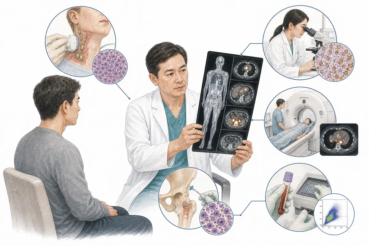
Basis for lymphoma diagnosis
1.Lymph node biopsy
Excision of an intact lymph node for pathological examination is the gold standard for diagnosing lymphoma; it identifies the histological type and immunohistochemical characteristics.
2.Immunohistochemical testing
Immunohistochemical analysis is performed on pathological specimens to detect markers such as CD20, CD3, CD30, and CD15, distinguishing between B-cell lymphoma, T-cell lymphoma, and Hodgkin lymphoma.
3.Whole-body CT scan
Contrast-enhanced CT assesses systemic lymph node involvement (cervical, mediastinal, abdominal, and retroperitoneal) and organ involvement, serving as a crucial tool for clinical staging.
4.PET-CT assessment
Whole-body PET-CT is a core tool for lymphoma staging; it can detect lesions invisible on conventional CT and guides precise staging and treatment response assessment.
5.Bone marrow aspiration and biopsy
Bone marrow aspiration and biopsy are used to assess the bone marrow for lymphoma cell infiltration, serving as a crucial component of staging and prognostic assessment.
6.Flow cytometry
Detection of lymphocyte immunophenotype in lymph node or bone marrow specimens to assist in differential diagnosis and clonality analysis.
7.Lactate dehydrogenase (LDH) test
Elevated serum LDH indicates a high tumor burden and is a key component of the International Prognostic Index (IPI) for lymphoma.
8.Genetic testing and analysis
Detection of gene rearrangements and mutations (e.g., MYC, BCL2, BCL6) assists in classification and prognostic evaluation, and guides targeted therapy.
9.EBV testing
Certain lymphomas (such as NK/T-cell lymphoma and Hodgkin lymphoma) are associated with Epstein-Barr virus (EBV) infection; EBER in situ hybridization aids in diagnosis.
Daily Self-Check
Seek medical attention promptly if painless lymph node enlargement is detected (especially if the diameter exceeds 1 cm and persists for more than 2 weeks).Avoid squeezing or applying heat to the lymph nodes.
Conclusion
Lymphoma diagnosis relies on lymph node biopsy and PET-CT staging.Pathological type and stage form the basis for developing personalized treatment plans.


