


Eye cancer primarily refers to retinoblastoma, the most common primary intraocular malignant tumor in infants and young children; it originates from retinal nuclear layer cells.The tumor can grow within the eye and spread outside the eyeball; in advanced stages, it may metastasize intracranially along the optic nerve or spread systemically via the bloodstream.The disease can occur in both hereditary and non-hereditary forms and may affect one or both eyes.
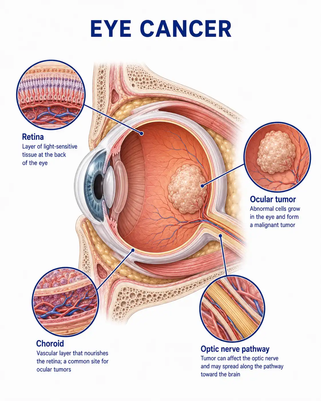
Global situation regarding eye cancer
Retinoblastoma has a global annual incidence of approximately 1 case per 15,000 to 20,000 live births, making it the most common intraocular malignancy in infants and young children.About two-thirds of cases are unilateral, while one-third are bilateral.Patients in developing countries are often diagnosed at an advanced stage, whereas early screening in developed countries has significantly increased the rate of eye preservation.
Consult a doctor regarding cancer-related issues
Parents should seek immediate ophthalmic consultation if they observe a yellowish-white reflection in the pupil (cat's-eye reflex), strabismus, redness or pain in the eye, or decreased vision in an infant or young child.Upon diagnosis, a treatment plan—whether eye-sparing or surgical—should be formulated under the guidance of an ophthalmic oncologist.
Main types of eye cancer
1.Unilateral non-hereditary type
Unilateral onset with no family history; typically presents between the ages of 2 and 5. The tumor affects only one eye, the prognosis is relatively favorable, and there is a high likelihood of preserving the eyeball.
2.Bilateral hereditary type
Bilateral disease (occurring sequentially or simultaneously) caused by a germline *RB1* gene mutation; onset typically occurs at an earlier age (often within the first year of life), necessitating lifelong monitoring for the risk of secondary tumors.
3.Trilateral retinoblastoma
Bilateral retinoblastoma accompanied by an intracranial ectopic retinoblastoma (located in the pineal or suprasellar region); a rare condition with a poor prognosis that requires multidisciplinary treatment.
4.Diffuse infiltrative type
A rare subtype characterized by diffuse, infiltrative growth along the retinal surface without forming a distinct mass; early diagnosis is challenging, and the tumor can rapidly spread along the optic nerve.
5.Recurrent/metastatic type
For advanced cases involving post-treatment recurrence or metastasis to the orbit, central nervous system, or other parts of the body, comprehensive salvage therapies—such as intensive chemotherapy, radiotherapy, and surgery—are required.
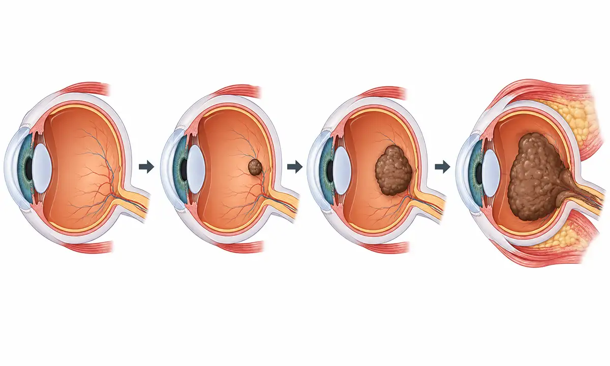
Typical symptoms and signs of eye cancer
1.Presentation of leukocoria (white pupil)
A yellowish-white or grayish-white reflection in the pupil area—commonly known as a "cat's-eye reflex"—is the most characteristic early sign; it is particularly noticeable in dim light or in photographs taken with a flash.
2.Appearance of strabismus
Tumor invasion of the macular region leads to vision loss; the affected child develops esotropia or exotropia, which is a common reason for parents to seek medical attention.
3.Red and painful eyes
Advanced-stage tumors can cause secondary glaucoma, leading to elevated intraocular pressure and symptoms such as ocular congestion, pain, photophobia, and lacrimation.
4.Exophthalmos
Extraocular extension of the tumor into the orbit causes exophthalmos or an orbital mass, indicating the disease has reached an advanced stage.
5.Decline in vision
When the tumor is located at the posterior pole or macular region, it can directly impair central vision; affected children may exhibit poor fixation, difficulty tracking objects, or significant vision loss.
6.Heterochromia iridis
Tumor seeding in the anterior chamber or iris neovascularization can lead to changes in iris color or abnormal pupil shape; hyphema or pseudohypopyon is occasionally observed.
What are the diagnostic methods for eye cancer
1.Fundus examination
Indirect ophthalmoscopy following pupil dilation is the primary diagnostic method; it allows for the observation of tumor size, location, number, and calcification, as well as the determination of the extent and stage of the lesion.
2.Ocular ultrasound
High-frequency B-mode ultrasound can visualize intraocular solid masses and calcifications, assess tumor size and location, and determine the presence of retinal detachment or vitreous seeding.
3.Orbital CT scan
CT can clearly visualize intratumoral calcifications and assess optic nerve thickening and orbital invasion, providing valuable information for staging and treatment planning.
4.Orbital MRI
MRI offers high soft-tissue resolution, clearly displaying the relationship between the tumor and surrounding tissues, and allows for the assessment of optic nerve invasion, intracranial extension, and lesions in the pineal region.
5.Systemic assessment examination
Includes bone marrow aspiration, lumbar puncture, and whole-body bone scanning to rule out central nervous system and systemic metastases and to determine disease stage.
6.Genetic testing
Detects *RB1* gene mutations to distinguish between heritable and non-heritable cases, guiding genetic counseling for the family, prenatal diagnosis, and monitoring for the risk of second primary tumors.
Eye cancer treatment methods
1.Local cryotherapy:Local cryotherapy involves directly freezing tumor tissue using a cryoprobe; it is suitable for small tumors located near the equator of the eye, allows for repeated treatments, and serves as a key local treatment method for eye preservation.
2.Laser photocoagulation:Laser photocoagulation (including transpupillary thermotherapy or direct laser destruction of the tumor) is suitable for small tumors in the posterior pole—particularly lesions outside the macular area—as it helps preserve central vision.
3.Systemic chemotherapy:Systemic chemotherapy can shrink tumors to facilitate local treatment; it is suitable for multiple tumors, vitreous seeding, and advanced cases.Commonly used drugs include carboplatin and vincristine.
4.Intra-arterial chemotherapy (via ophthalmic artery infusion): Chemotherapy drugs are delivered directly to the tumor site at high concentrations; this method improves eye-preservation rates and reduces systemic side effects, making it an important option for intermediate-stage cases.
5.Radiotherapy: Local scleral plaque radiotherapy or external beam radiotherapy; suitable for tumors that are insensitive to chemotherapy or have recurred.Use with caution in patients with hereditary forms, as it increases the risk of secondary tumors.
6.Enucleation: Enucleation is required when the tumor is large, there is no hope of preserving vision, or the optic nerve has been invaded.Postoperative pathological examination clarifies the stage and guides subsequent adjuvant therapy.
7.Orbital exenteration: If the tumor has invaded the orbit or the optic nerve stump tests positive, orbital exenteration is required; postoperative comprehensive treatment combining radiotherapy and chemotherapy is used to control local disease.
8.Follow-up monitoring: Regular ophthalmological examinations to monitor the contralateral eye and residual lesions.Patients with hereditary forms require lifelong follow-up to monitor for the development of second malignancies, such as osteosarcoma and melanoma.
Conclusion
Retinoblastoma is the most common intraocular malignancy in infants and young children, and early detection and treatment are key to preserving the eye and vision.With the development of comprehensive treatment modalities—such as local therapy, chemotherapy, and intra-arterial infusion—eye preservation rates have risen significantly, and prognoses continue to improve.


