


The global annual incidence of retinoblastoma is approximately one case per 15,000 to 20,000 live births.About 40% of cases are hereditary and associated with *RB1* gene mutations.With early detection, over 95% of affected children survive, and more than 50% retain their vision;however, in developing countries, the disease is often at an advanced stage by the time of diagnosis.
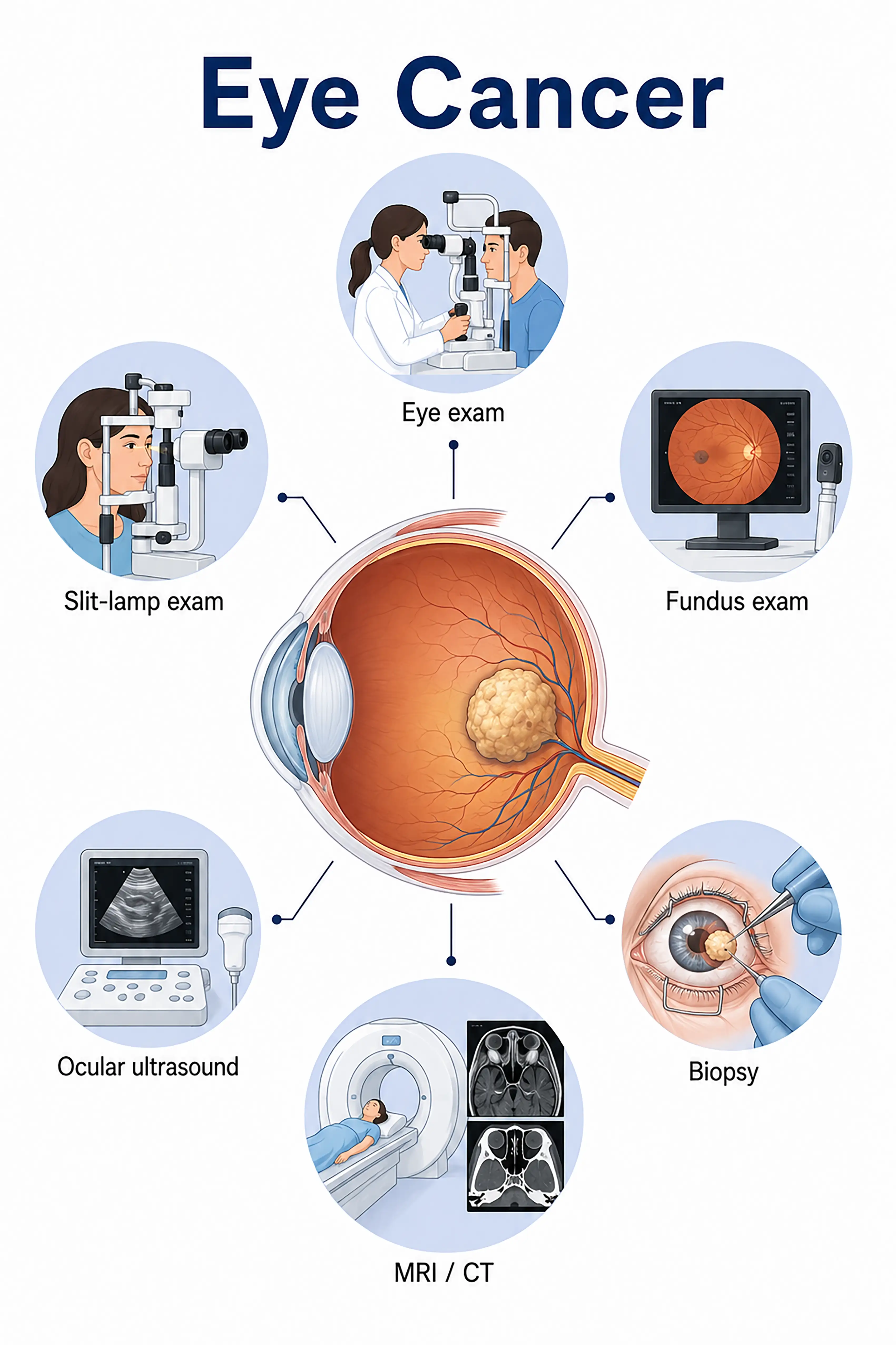
Diagnosis based on eye cancer stage
1.Early diagnosis of eye cancer
Fundus examination reveals a white or grayish-white mass on the retina.Indirect ophthalmoscopy following pupil dilation is the primary diagnostic method, allowing for the determination of the tumor's number, location, and size.
2.Diagnosis of intermediate-stage eye cancer
Leukocoria (white pupil), strabismus, or eye redness.Ocular ultrasound reveals a solid intravitreal mass with calcification, and orbital CT shows a high-density calcified lesion; the diagnosis is confirmed by correlating these findings with the fundus appearance.
3.Diagnosis of advanced eye cancer
The tumor extends extraocularly into the orbit or metastasizes distantly.MRI is used to assess optic nerve and intracranial invasion, while whole-body bone scans and bone marrow examinations rule out metastasis and clarify the stage.
If eye cancer is not diagnosed promptly, the tumor may spread intracranially along the optic nerve or metastasize via the bloodstream to the bones and bone marrow, endangering life and necessitating enucleation (removal of the eyeball).
When to Seek Medical Attention
Early signs of eye cancer include a whitish appearance in the pupil area of infants and young children (leukocoria), strabismus (squinting), eyeball protrusion, and eye redness.Please consult a doctor via the "Chat with a Doctor" feature to facilitate early detection.During the online consultation, the doctor will ask about the eye abnormalities you have observed and guide you through a fundus examination.
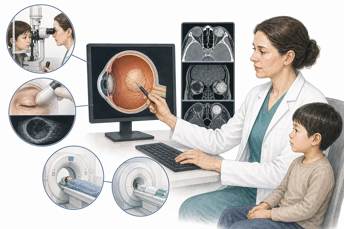
Basis for diagnosing eye cancer
1.Fundus examination
Indirect ophthalmoscopy following pupil dilation is the core method for diagnosing retinoblastoma, allowing for direct visualization of the tumor's location, size, number, and calcification on the retina.
2.Ocular ultrasound examination
High-frequency ocular B-mode ultrasound can visualize solid intravitreal masses, calcifications, and retinal detachment, as well as assess tumor size and location; it is particularly useful in cases with opacities of the refractive media.
3.Orbital CT scan
CT can clearly visualize intratumoral calcifications—making it the most sensitive method for detecting calcification—while also allowing for the assessment of orbital bone and the optic canal.
4.Orbital MRI examination
MRI assesses whether the tumor invades the optic nerve or orbital soft tissues or extends intracranially; as it involves no radiation, it is suitable for examination and follow-up in infants and young children.
5.Fundus fluorescein angiography
FFA reveals tumor vascular characteristics and aids in the differential diagnosis of other causes of leukocoria (such as Coats' disease and persistent hyperplastic primary vitreous).
6.Examination under general anesthesia
Perform a detailed fundus examination and fundus photography under general anesthesia to comprehensively assess and document the status of tumors in both eyes.
7.Genetic testing
Detects *RB1* gene mutations to distinguish between heritable and non-heritable forms; heritable cases require lifelong follow-up and monitoring for the risk of second primary tumors.
8.Lumbar puncture
When optic nerve invasion or intracranial metastasis is suspected, cerebrospinal fluid is sampled to check for tumor cells and assess central nervous system involvement.
9.Systemic assessment
Bone marrow aspiration and whole-body bone scanning are used to rule out distant metastasis; pediatric patients with genetic forms require lifelong monitoring for secondary tumors (such as osteosarcoma or melanoma).
Daily Self-Check
Parents should seek immediate medical attention if they notice a white pupil or strabismus in an infant or young child.Those with a family history should undergo regular fundus screening after birth.
Conclusion
The diagnosis of retinoblastoma relies on fundus examination and imaging.Early detection can preserve the eyeball and even vision, whereas delayed diagnosis can lead to blindness and life-threatening consequences.


