


Ependymoma is a tumor formed by the abnormal proliferation of ependymal cells.Ependymal cells line the walls of the ventricles and the inner surface of the spinal cord's central canal, playing a role in the production and circulation of cerebrospinal fluid.
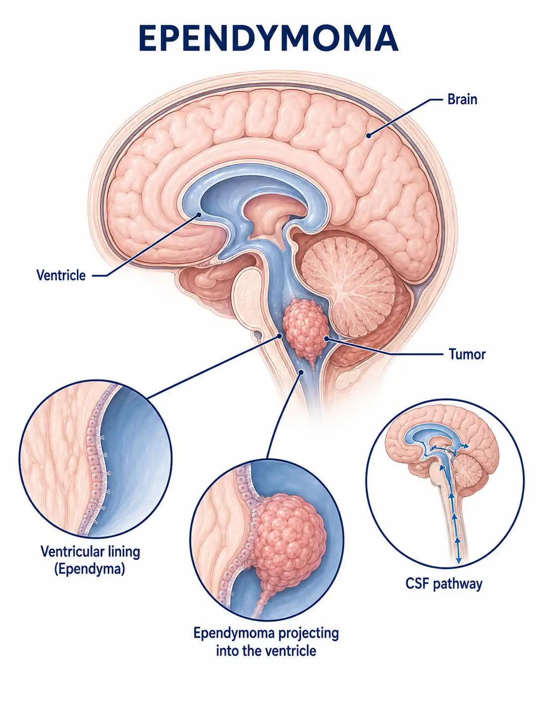
Global overview of ependymoma
Ependymomas account for approximately 5%–6% of all intracranial gliomas; about 69% occur in children, making them one of the more common posterior fossa tumors in the pediatric population.In adults, they are most frequently found in the spinal cord, with no significant difference in incidence between males and females.
Consult a doctor regarding cancer-related issues
Children or adults experiencing symptoms such as headache and vomiting, neck stiffness, unsteady gait, or back pain accompanied by limb weakness should promptly consult a neurosurgeon.
Main types of ependymoma
1. Myxopapillary ependymoma
WHO Grade I; occurs almost exclusively in the conus medullaris and cauda equina regions; slow-growing with a favorable prognosis.
2. Subependymoma
WHO Grade I; slow-growing; typically located on the ventricular wall; composed of clusters of tumor cells embedded in a fibrous matrix;prognosis is favorable.
3. Ependymoma
WHO Grade II; can occur intracranially or in the spinal cord; includes subtypes such as papillary, clear cell, and tanycytic.
4. Anaplastic ependymoma
WHO Grade III; characterized by active cell proliferation, accompanied by vascular proliferation and necrosis; highly malignant with a tendency for recurrence.
5. RELA fusion-positive
Characterized by the presence of the RELA fusion gene and WHO grade II–III status; primarily observed in pediatric supratentorial tumors and associated with a relatively poor prognosis.
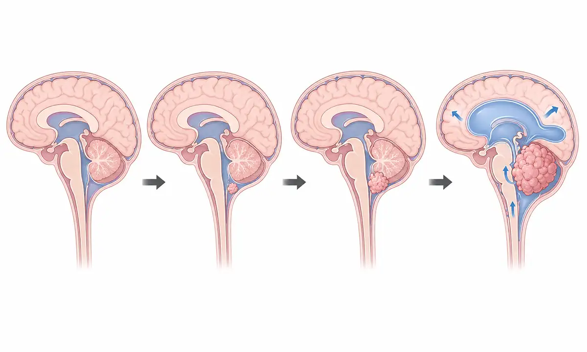
Typical symptoms and signs of ependymoma
1. Headache and vomiting
Obstruction of cerebrospinal fluid circulation by the tumor causes obstructive hydrocephalus, leading to headache, nausea, and vomiting; symptoms are most pronounced in the morning.
2. Neck stiffness
When a posterior fossa tumor extends inferiorly into the foramen magnum region, symptoms such as neck pain, stiffness, and torticollis may occur, worsening with movement.
3. Unsteady gait
Ataxia occurs when the tumor invades the cerebellum, manifesting as a swaying walk, unsteady gait, and a tendency to fall.
4. Visual acuity changes
Increased intracranial pressure causes papilledema, leading to reduced visual acuity, while tumor compression of the visual pathways can result in visual field defects.
5. Back pain
Spinal cord ependymomas often manifest as back or neck pain radiating to the limbs, worsening at night, and accompanied by sensory abnormalities.
6. Limb weakness
Compression of the spinal cord by an ependymoma causes limb weakness, numbness, and bowel/bladder dysfunction; severe cases can lead to paralysis.
Diagnostic Methods for Ependymoma
1. Neurological examination
A detailed assessment of cranial nerve function, muscle strength, sensation, reflexes, and coordination helps localize the lesion.
2. Cranial MRI scan
MRI is the preferred method for diagnosing ependymomas; typical findings include a space-occupying lesion at the floor of the fourth ventricle, characterized by heterogeneous enhancement and irregular morphology.
3. Whole-spine MRI
Plain and contrast-enhanced scans of the entire spinal cord help detect potential lesions resulting from cerebrospinal fluid dissemination and are crucial for formulating treatment plans.
4. Pathological biopsy
Pathological examination following surgical tumor resection is the gold standard for diagnosis and grading; the condition typically presents with rosette-like structures.
5. Cerebrospinal fluid examination
Lumbar puncture is performed to obtain CSF for cytological examination; a positive result indicates tumor dissemination via the cerebrospinal fluid.
6. Genetic testing
Detects *RELA* fusion genes and other molecular markers, aiding in subtype identification and treatment guidance.
Ependymoma Treatment Methods
1. Surgical treatment
Complete surgical resection is the primary treatment for ependymoma; the extent of resection is the most critical prognostic factor, requiring meticulous technique to protect surrounding structures.
2. Radiation therapy
Postoperative adjuvant radiotherapy is indicated for anaplastic and incompletely resected ependymomas; craniospinal irradiation is used for disseminated disease.
3. Chemotherapy
Chemotherapy has limited efficacy and is primarily used for infants and young children to delay radiotherapy or for patients with recurrent disease;regimens based on lomustine are commonly employed.
4. Targeted therapy
Drugs targeting specific molecular markers are under investigation, offering new therapeutic possibilities for patients with recurrent or refractory ependymoma.
5. Cerebrospinal fluid shunting
For patients with severe hydrocephalus and unresectable tumors, a ventriculoperitoneal shunt is performed first to alleviate symptoms of increased intracranial pressure.
6. Symptomatic and supportive care
Glucocorticoids alleviate cerebral edema and symptoms, antiemetics control nausea and vomiting, and nutritional support improves general health status.
7. Rehabilitation therapy
Postoperative physical therapy and functional training improve motor function and activities of daily living, thereby enhancing quality of life.
8. Follow-up monitoring
Regular postoperative whole-brain and whole-spine MRI follow-up monitors for local recurrence and CSF dissemination; for pediatric patients, growth and development require attention.
Conclusion
Ependymoma is a nervous system tumor originating from ependymal cells, most commonly occurring in children and the spinal cord region.Striving for complete surgical resection, combined with necessary adjuvant therapy, helps improve the patient's prognosis.


