


Ependymomas account for approximately 2%–5% of all central nervous system tumors.They can occur at any age but are relatively common in children, representing a frequent type of pediatric intracranial tumor.They are classified as WHO Grade I–III; spinal cord ependymomas are more common in adults.
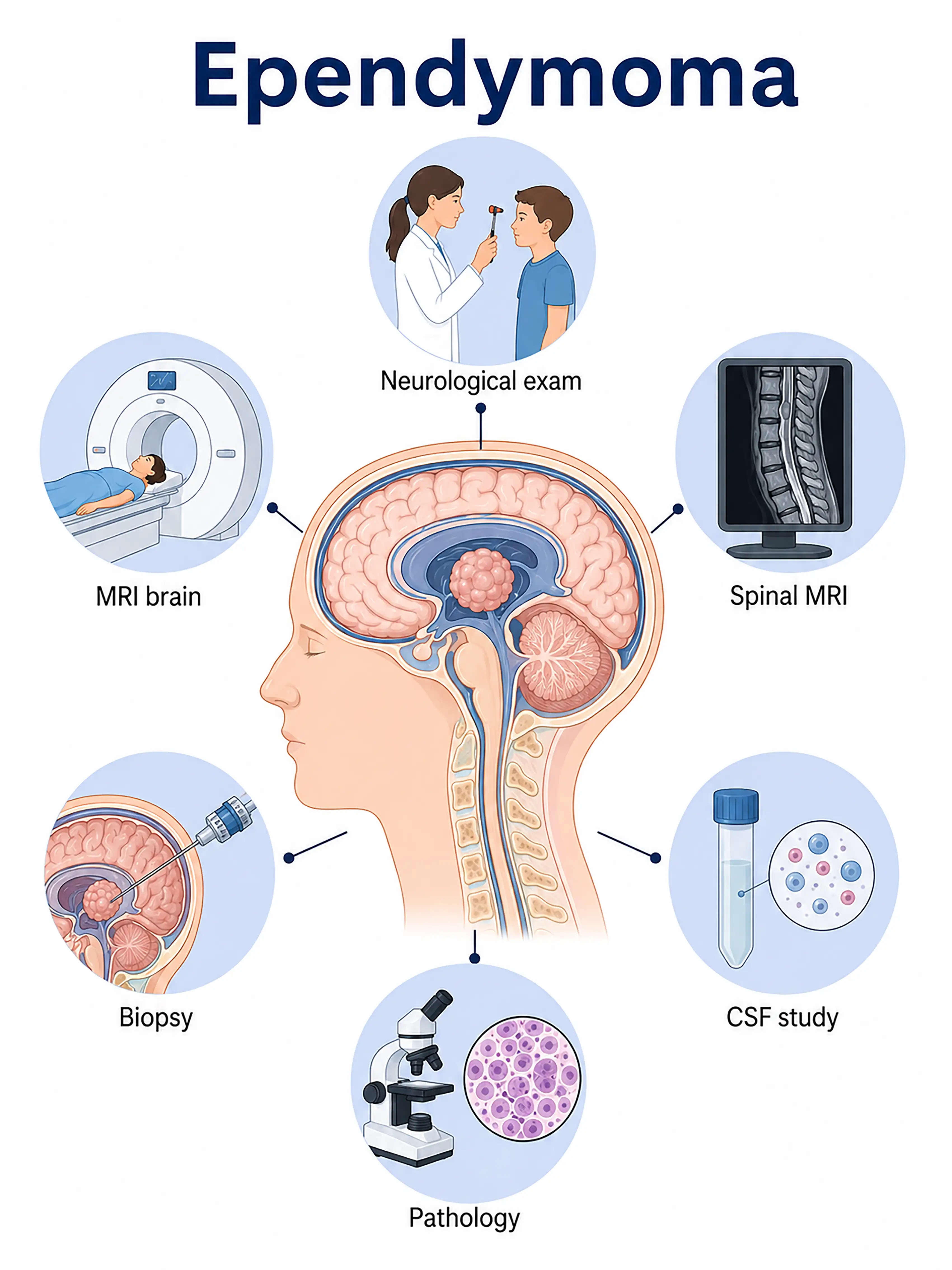
Diagnosis based on ependymoma staging
1.Early diagnosis of ependymoma
The tumor is small, slow-growing, and often asymptomatic.MRI reveals a well-defined intraventricular or intramedullary lesion that is isointense or hypointense on T1-weighted images, hyperintense on T2-weighted images, and shows marked enhancement after contrast administration.
2.Diagnosis of intermediate-stage ependymoma
Symptoms of hydrocephalus (headache, vomiting) or spinal cord compression (limb numbness, weakness) appear.MRI shows tumor enlargement with peritumoral edema; surgical sampling is required to determine the pathological grade.
3.Diagnosis of advanced ependymoma
Severe hydrocephalus causes a rapid rise in intracranial pressure, or spinal cord compression leads to paraplegia.Surgical decompression and specimen acquisition are performed to determine the WHO grade and molecular characteristics.
If not diagnosed promptly, intracranial ependymomas can lead to fatal hydrocephalus and brain herniation, while spinal cord ependymomas can result in permanent paraplegia and bowel or bladder incontinence.
When to Seek Medical Attention
Early signs of ependymoma include persistent headaches, difficulty walking, neck or back pain, and limb numbness.Please consult a doctor via the "Chat with a Doctor" feature for early detection.During the online consultation, the doctor will ask about your symptoms and disease progression, and guide you through the process of obtaining an MRI scan.
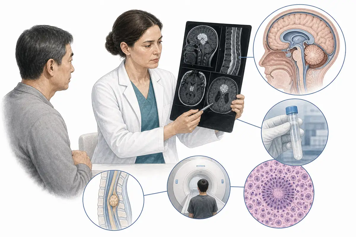
Basis for Ependymoma Diagnosis
1.Cranial/spinal MRI
MRI is the preferred method for diagnosing ependymomas.Typical findings include a well-defined mass within the ventricles or the central canal of the spinal cord, appearing isointense or hypointense on T1-weighted images and hyperintense on T2-weighted images, with marked, homogeneous enhancement after contrast administration.
2.Surgical histopathology
Pathological examination of tissue specimens obtained via surgical resection is the gold standard for diagnosing ependymoma and allows for the determination of the WHO grade (Grades I–III).
3.CT scan
CT can reveal intraventricular masses of isodensity or hyperdensity (appearing hyperdense when calcified) and allows for rapid assessment of hydrocephalus and the degree of ventricular enlargement.
4.Cerebrospinal fluid examination
Lumbar puncture is performed to obtain CSF for cytological examination to look for shed tumor cells and assess for the presence of CSF dissemination.
5.RELA fusion gene testing
Detects the *RELA* fusion gene (*RELA-FIP1L1*), a key molecular marker for pediatric supratentorial ependymoma that is associated with prognosis.
6.NF2 gene mutation testing
Detection of NF2 gene mutations aids in differentiating certain ependymoma subtypes, particularly those associated with neurofibromatosis type 2.
7.Myelography
Conventional myelography can reveal filling defects caused by intramedullary space-occupying lesions, but it has been superseded by MRI and is used only in special circumstances.
8.Intraoperative neurophysiological monitoring
Real-time intraoperative monitoring of neurological function protects the spinal cord and vital functional areas of the brainstem, enhancing surgical safety.
9.Postoperative MRI follow-up
Regular postoperative MRI examinations assess the extent of tumor resection, the presence of recurrence, and changes in syringomyelia; this is the core method for long-term follow-up.
Daily Self-Check
Seek medical attention promptly if unexplained headache, difficulty walking, or back pain occurs.Individuals with a family history of neurofibromatosis should undergo regular neurological examinations.
Conclusion
Diagnosis relies primarily on MRI and surgical pathology.Complete surgical resection is key to a cure, while molecular testing aids in classification and prognostic assessment.


