


Endometrial cancer is a common female reproductive malignancy worldwide, and incidence is rising in many regions. It mainly affects postmenopausal women; obesity, metabolic disorders, and prolonged estrogen exposure increase risk. Many patients are diagnosed early and have a relatively favorable prognosis.
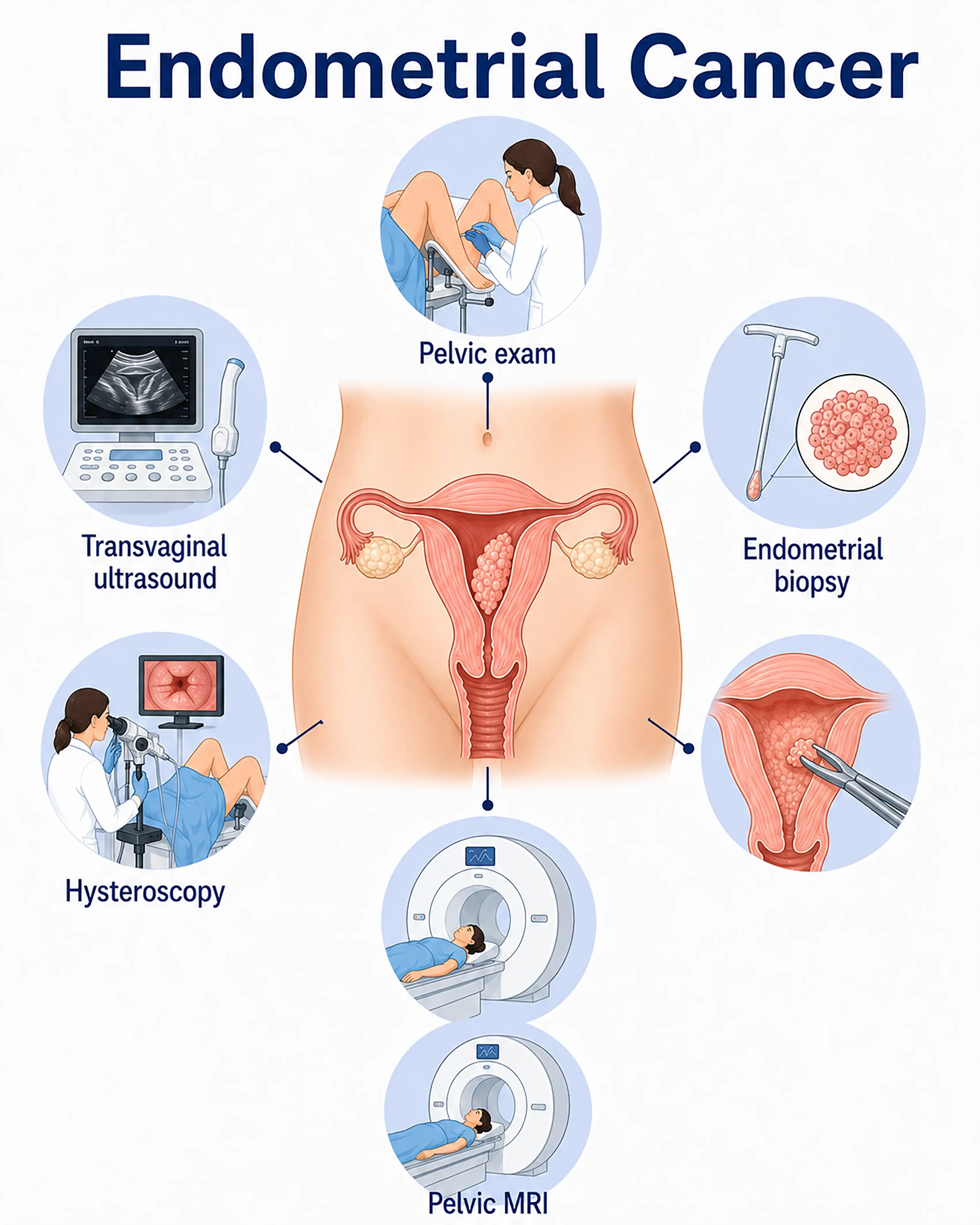
Endometrial cancer diagnosis by stage
1.Early-stage endometrial cancer diagnosis
The tumor remains within the endometrium, usually causing postmenopausal bleeding or menstrual irregularities. Transvaginal ultrasound shows endometrial thickening greater than 5 mm, requiring biopsy for definitive diagnosis.
2.Intermediate-stage endometrial cancer diagnosis
The tumor invades the myometrium. Ultrasound and MRI show endometrial thickening with myometrial invasion. Fractional curettage obtains tissue for pathology to identify histological type and grade.
3.Advanced-stage endometrial cancer diagnosis
The tumor invades the cervix or adnexa or spreads distantly. CT or MRI assesses metastatic extent, surgical exploration establishes stage, and pathology guides subsequent treatment.
Without timely diagnosis, the tumor continues to infiltrate the myometrium and spread nearby, increasing distant metastasis risk and compromising treatment outcomes and prognosis.
When to Seek Medical Attention
Early warning signs include postmenopausal vaginal bleeding, markedly heavier or prolonged periods, and abnormal vaginal discharge. Please consult a doctor through "Chat with a Doctor" to support early detection. The doctor will ask about menstrual history and symptoms and guide you through ultrasound and endometrial biopsy.
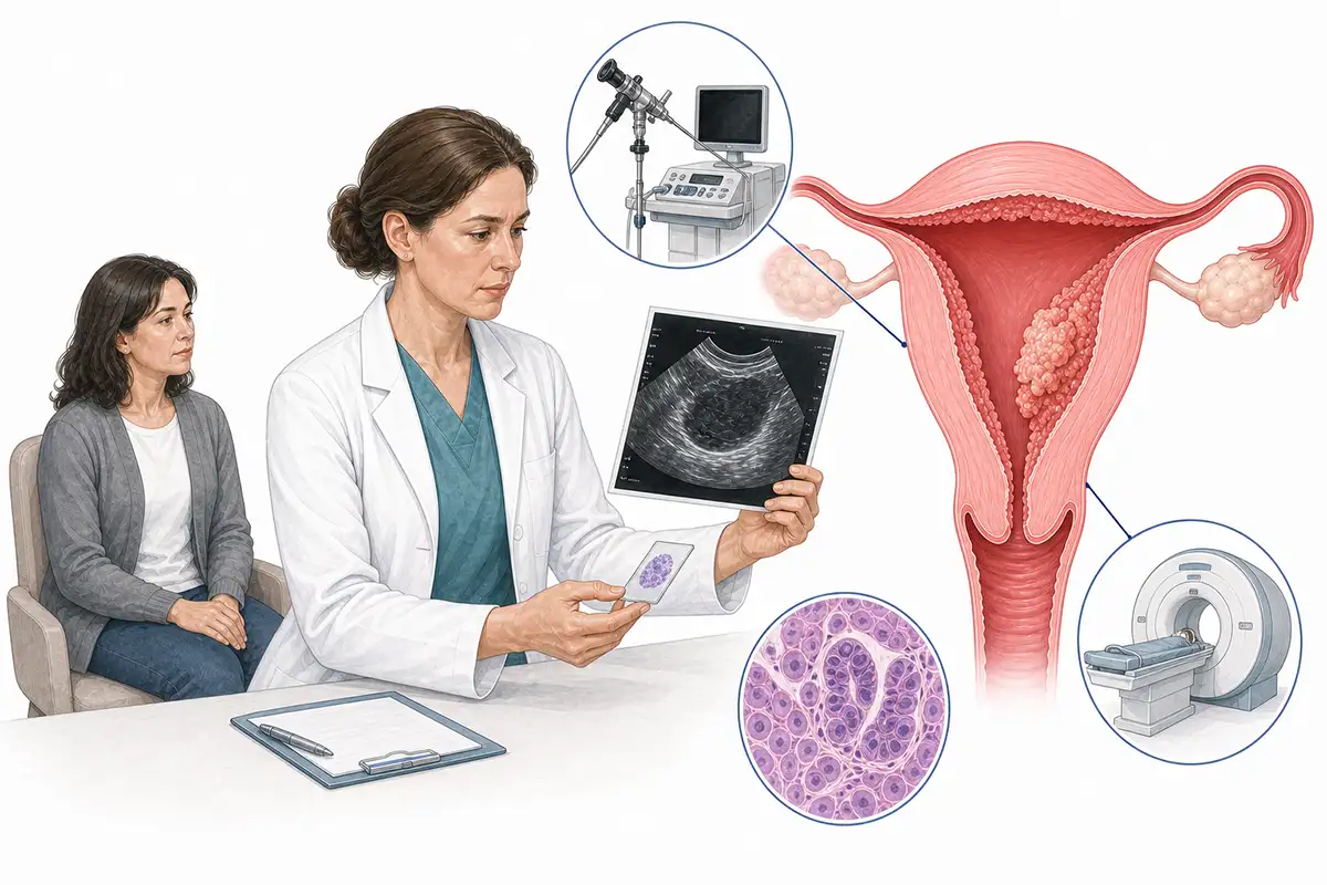
Basis for endometrial cancer diagnosis
1.Transvaginal ultrasound
This preferred noninvasive examination measures endometrial thickness and identifies intrauterine masses. Postmenopausal thickness greater than 5 mm warrants concern.
2.Endometrial biopsy
Suction or a curette obtains endometrial tissue for pathology. This principal diagnostic method can be performed in an outpatient setting.
3.Diagnostic curettage
Fractional curettage separately samples the cervical canal and uterine cavity lining to establish disease location and extent. It is a traditional diagnostic method.
4.Hysteroscopy
A hysteroscope is passed through the vagina into the uterine cavity for direct inspection and precisely targeted biopsy, improving detection of early lesions.
5.Pelvic MRI
MRI clearly shows endometrial thickness, myometrial invasion depth, and cervical involvement and is valuable for preoperative staging.
6.Tumor marker testing
Serum CA125 elevation correlates with disease extent and stage and can be used for preoperative assessment and postoperative follow-up.
7.Chest and abdominal CT
Assessment of distant metastases in the lungs, liver, and retroperitoneal nodes establishes stage and guides treatment selection.
8.Histopathological examination
Pathology of the surgically obtained uterine specimen establishes histological type, grade, and myometrial invasion depth and is the diagnostic gold standard.
9.Genetic counseling and screening
Testing young patients and those with a family history for Lynch syndrome-related genes such as MLH1 and MSH2 helps assess hereditary risk and develop follow-up plans.
Daily Self-Check
Postmenopausal vaginal bleeding requires immediate medical attention. Markedly heavier or prolonged periods in reproductive-age women also warrant concern. Regular gynecological examinations and ultrasound screening support early detection.
Conclusion
Endometrial cancer has a relatively high early diagnosis rate, with postmenopausal bleeding the most important warning sign. Transvaginal ultrasound with endometrial biopsy is central to diagnosis, and early treatment has a favorable prognosis.


