


Duodenal cancer is the most common type of malignant small bowel tumor, accounting for approximately 40%–50% of cases.It has a low overall incidence and is classified as a rare tumor.It predominantly affects individuals over the age of 50 and is associated with familial adenomatous polyposis (FAP) and Crohn's disease.Early symptoms are often atypical, making diagnosis difficult.
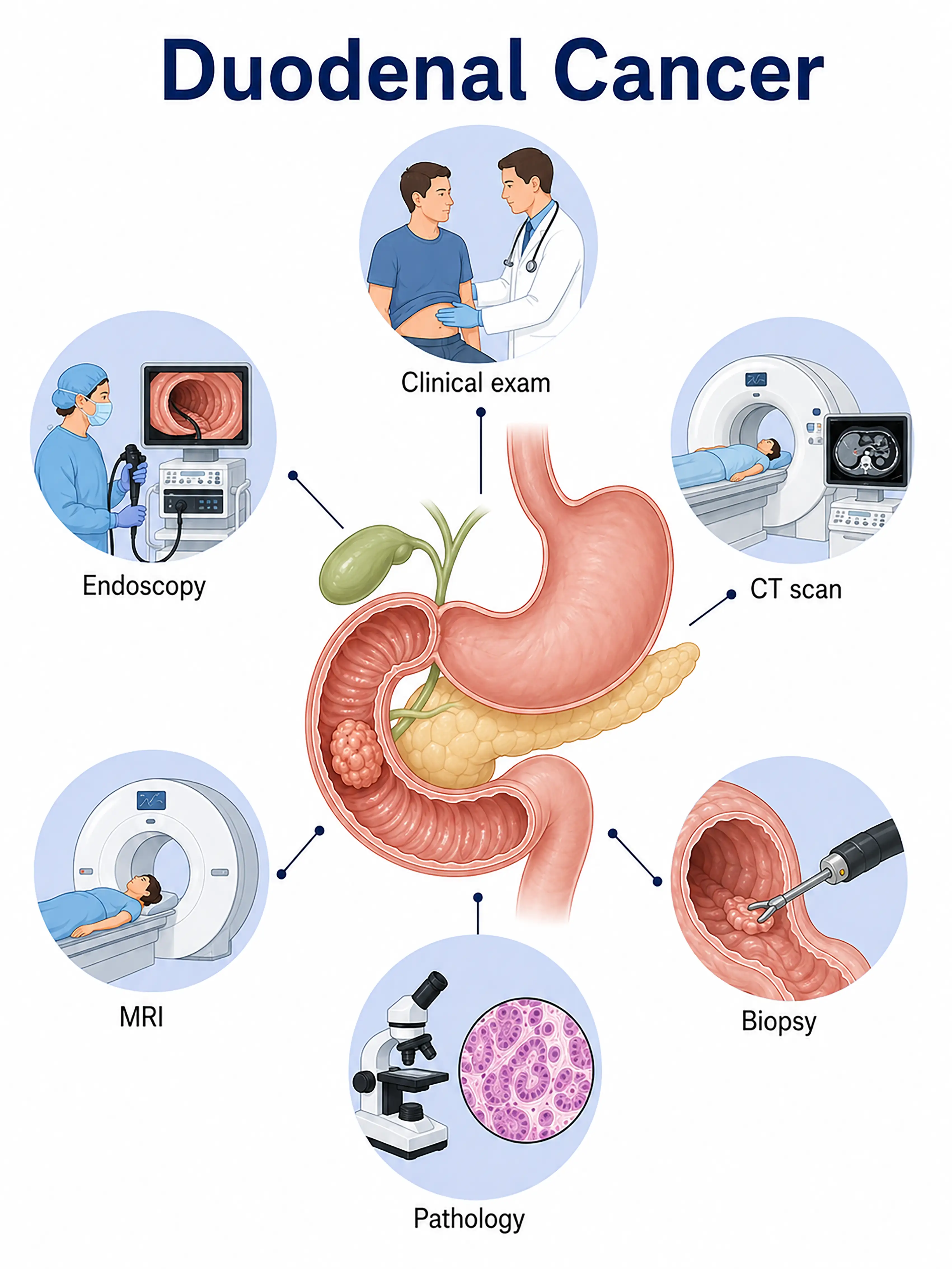
Diagnosis of duodenal cancer stages
1.Early diagnosis of duodenal cancer
The tumor is confined to the duodenal mucosa; there are usually no obvious symptoms.Lesions are detected via gastroscopy in the descending duodenum or around the duodenal papilla, and the diagnosis is confirmed by biopsy.
2.Diagnosis of intermediate-stage duodenal cancer
Symptoms include upper abdominal pain, jaundice, or gastrointestinal bleeding.Gastroscopy and endoscopic ultrasound (EUS) are used to assess the depth of tumor invasion and regional lymph node metastasis, while biopsy confirms the pathological type.
3.Diagnosis of advanced duodenal cancer
The tumor invades the pancreas or metastasizes distantly to the liver or lungs.Contrast-enhanced CT and MRI are used to assess local invasion and the extent of metastasis, while surgical exploration determines the stage.
If duodenal cancer is not diagnosed promptly, the tumor may invade vital organs such as the pancreas, making surgery extremely difficult and resulting in a poor prognosis.
When to Seek Medical Attention
Early signs of duodenal cancer include upper abdominal pain, jaundice, and melena (black, tarry stools).Please consult a doctor via the "Chat with a Doctor" feature for early detection.During the online consultation, the doctor will ask about your symptoms and medical history and guide you through procedures such as gastroscopy and abdominal CT scans.
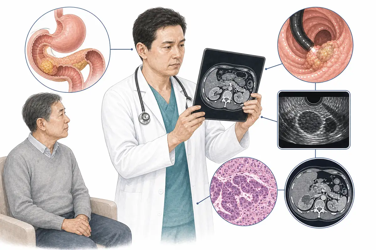
Basis for diagnosing duodenal cancer
1.Gastroscopy and biopsy
Gastroscopy is the preferred method for diagnosing duodenal cancer; it enables direct visualization of lesions in the descending duodenum and the peri-ampullary region, as well as the collection of tissue for pathological examination, serving as the gold standard for diagnosis.
2.Endoscopic ultrasound examination
EUS clearly visualizes the layers of the duodenal wall and the depth of tumor invasion, assesses pancreaticobiliary involvement and regional lymph node metastasis, and enables precise T-staging.
3.Abdominal CT scan
Contrast-enhanced CT evaluates tumor size, its relationship with surrounding blood vessels and the pancreas, and the presence of abdominal lymph node and liver metastases, guiding the assessment of surgical feasibility.
4.Abdominal MRI
MRI offers high soft-tissue resolution, allowing for the assessment of the tumor's relationship with pancreatic and bile ducts and the visualization of hepatic metastases, which is crucial for surgical planning.
5.Tumor marker test
Measurement of serum CEA and CA19-9 levels; although specificity is limited, elevated levels indicate a need for further examination and serve as tools for monitoring treatment efficacy and follow-up.
6.Duodenal hypotonic radiography
X-ray examination following oral barium administration reveals destruction of the duodenal mucosa, filling defects, and luminal narrowing; allows for observation of the extent of the lesion and intestinal motility.
7.ERCP examination
Endoscopic retrograde cholangiopancreatography (ERCP) can visualize compression or obstruction of the bile and pancreatic ducts and allows for the collection of pancreatic juice or bile duct brushings for cytologic examination.
8.Capsule endoscopy
For lesions in the distal duodenum that are difficult to reach via gastroscopy, capsule endoscopy can provide supplementary imaging information to detect mucosal lesions.
9.Laparoscopic exploration
For cases where imaging findings are inconclusive, laparoscopic exploration allows for direct visualization to assess tumor extent and the presence of abdominal metastases, as well as the acquisition of tissue biopsies.
Daily Self-Check
Seek medical attention promptly if persistent upper abdominal pain, jaundice, or melena occurs.Individuals with a history of familial adenomatous polyposis or Crohn's disease should undergo regular gastrointestinal examinations.
Conclusion
Duodenal cancer is relatively rare, and early symptoms are often atypical.Gastroscopy combined with endoscopic ultrasound is a key diagnostic tool, and early detection is crucial for successful surgical resection.


