


Global disease prevalence: Craniopharyngioma is a rare intracranial tumor worldwide; it is relatively more common in children and adolescents. Although benign, its deep location makes treatment challenging.
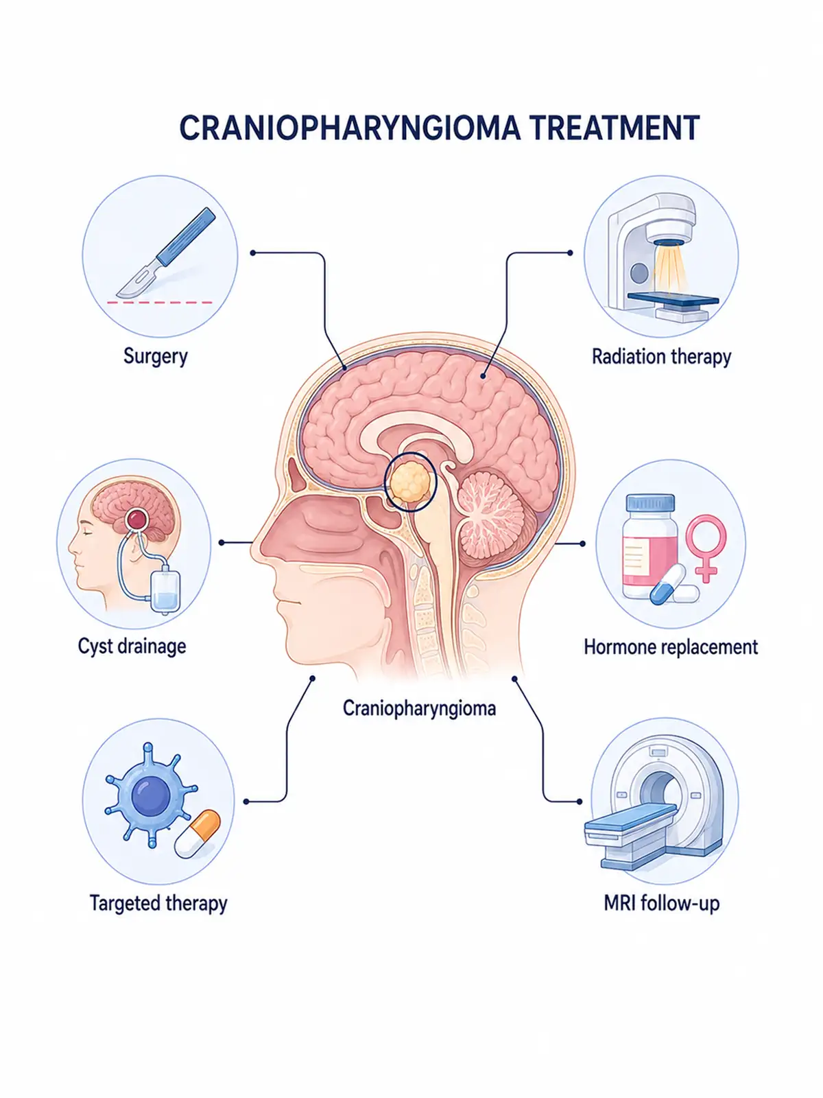
Staged treatment for craniopharyngioma
Early treatment (involving a localized, small tumor with a favorable response)
specifically surgical resection combined with adjuvant therapy—can effectively control tumor progression, and the patient's prognosis is relatively good.
Advanced-stage treatment (addressing severe compression, alleviating symptoms, and prolonging survival)
primarily employs systemic therapies such as radiotherapy to relieve symptoms of neurological compression and maximize patient survival time.
Consequences of delayed early treatment: Failure to receive timely, standardized treatment in the early stages allows the tumor to continue enlarging and compressing the hypothalamus and optic nerves; missing the optimal window for intervention leads to a worsening of the condition.
Why choose Fosun Health for craniopharyngioma treatment
Leveraging the neurosurgery, M6 CyberKnife, VitalBeam, ophthalmology, endocrinology, and imaging platforms of its affiliated hospitals, Fosun Health facilitates multidisciplinary consultations involving experts in neurosurgery, radiotherapy, ophthalmology, endocrinology, medical imaging, and rehabilitation.Based on tumor size, cystic-solid structure, and the tumor's relationship with the visual pathway, hypothalamus, and pituitary stalk, the team determines the treatment strategy, such as total resection or subtotal resection combined with radiotherapy.
Endoscopic surgery and precision radiotherapy help minimize impact on the optic nerve, hypothalamus, pituitary gland, and normal brain tissue, thereby reducing the risk of adverse effects related to vision, endocrine function, and cognition.Fosun Health provides comprehensive, long-term medical support covering everything from diagnosis and treatment to hormone replacement, fluid and electrolyte balance, weight management, sleep hygiene, and cognitive care.

Treatment Methods for Craniopharyngioma
1.M6 CyberKnife treatment
Indicated for small-volume solid residual or recurrent lesions confirmed by MDT, where the distance between the lesion and the optic nerve/optic chiasm meets dose constraint requirements.
Treatment advantages:
① Targeting small residual volumes: Reduces radiation exposure to normal tissue in the sellar region.
② Hypothalamus protection: Reduces impact on body weight, metabolism, and cognitive function.
③ Hypofractionated treatment: Offers an option for patients who cannot undergo further surgery.
④ Strict requirements regarding the visual pathway: Generally unsuitable when in close proximity to the optic nerve or optic chiasm.
2.VitalBeam precision radiotherapy
Indicated for craniopharyngiomas that are residual post-surgery, recurrent, or unsuitable for complete resection due to risks involving the visual pathways or hypothalamus.
Treatment advantages:
① Covers cystic-solid residual disease: Suitable for irregular surgical cavities and adjacent areas.
② Protect optic nerves: Control the single-fraction dose to visual pathways through fractionated therapy.
③ Protects the hypothalamus and pituitary gland: Reduces radiation exposure to critical endocrine structures.
④ Supports a subtotal resection strategy: Controls residual tumor while preserving function.
3. Endoscopic transnasal or microsurgical resection and decompression
Suitable for patients presenting with visual pathway compression, hydrocephalus, or cystic expansion, or those requiring definitive pathological diagnosis.
4.Intracystic chemotherapy
Chemotherapy drugs are injected directly into the tumor cyst cavity to precisely kill the tumor cells within;this method is suitable for patients with cystic craniopharyngioma.
5.Targeted therapy
Precision targeting of specific markers, such as the BRAF mutation, offers an effective treatment option for some patients with craniopharyngioma.
6.Proton beam therapy
Uses proton beams to precisely target tumor tissue while maximizing protection of surrounding normal tissue; suitable for pediatric patients with craniopharyngioma.
Post-treatment care
Regularly monitor hormone levels and changes in vision; maintain a regular daily routine; attend scheduled follow-ups and actively cooperate with further treatment as advised.
Post-treatment examinations
After treatment, regular cranial MRI scans, hormone level testing, and vision checks are required to detect tumor recurrence or changes in the condition in a timely manner.
Conclusion
Although craniopharyngioma is benign, the risks it poses cannot be ignored; the key to treatment lies in early detection, diagnosis, and intervention, as well as selecting an appropriate treatment plan and adhering to follow-up care.


