


Craniopharyngiomas account for approximately 2%–5% of intracranial tumors and are the most common sellar region tumors in children.There are two peak incidence periods: 5–14 years and 50–74 years.Although benign, their deep location makes surgical treatment challenging and recurrence rates high.
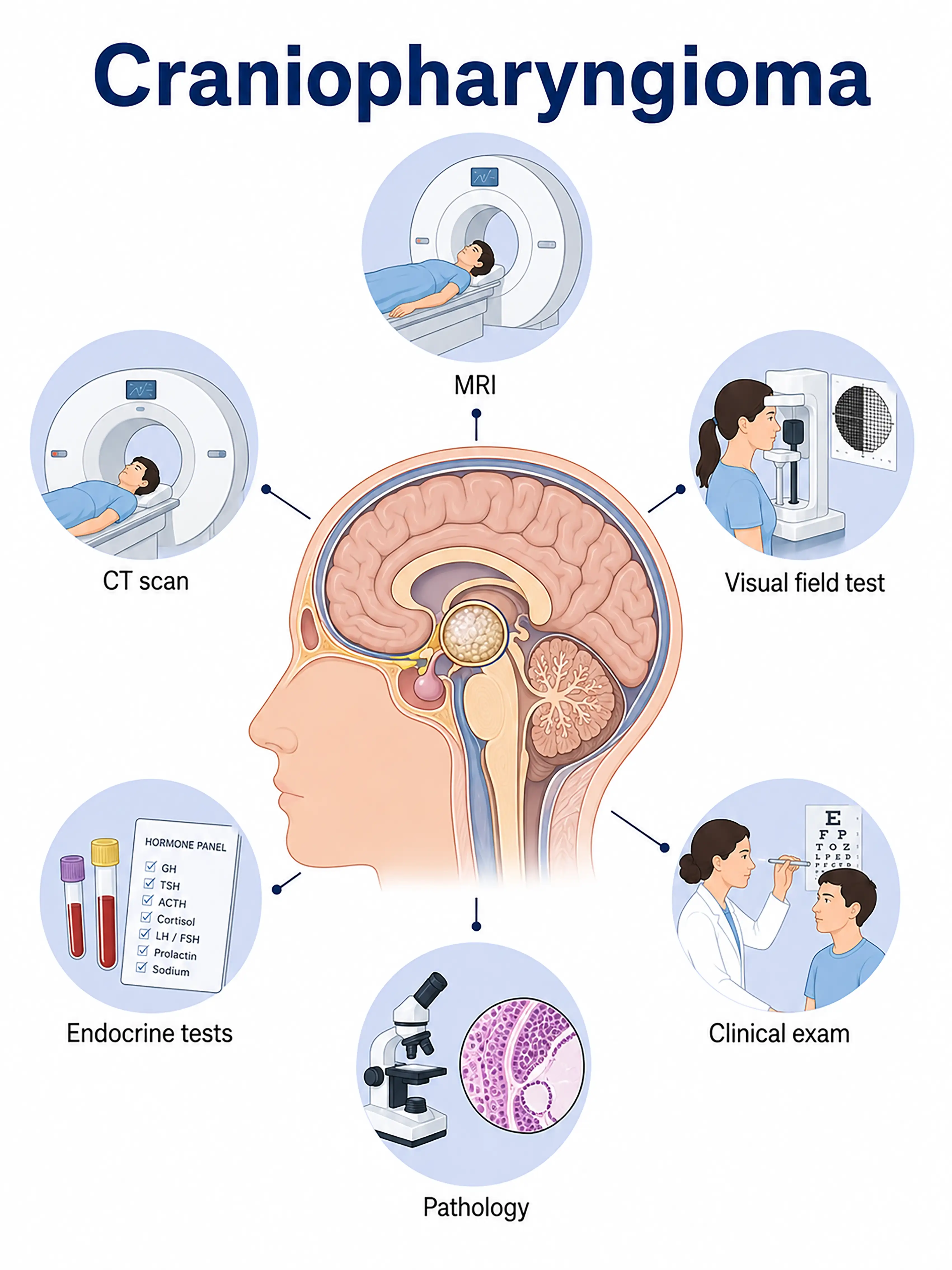
Diagnosis based on craniopharyngioma stages
1.Early diagnosis of craniopharyngioma
Small tumors compressing the pituitary gland or hypothalamus.MRI shows a suprasellar cystic-solid lesion with high signal intensity on T1WI (due to cholesterol crystals) and T2WI, accompanied by calcification; contrast enhancement reveals enhancement of the cyst wall.
2.Diagnosis of intermediate-stage craniopharyngioma
Symptoms include changes in vision or visual fields, diabetes insipidus, and stunted growth or developmental delay.MRI scans show tumor enlargement compressing the optic chiasm and the third ventricle; endocrine testing is used to assess pituitary function.
3.Diagnosis of advanced craniopharyngioma
A large tumor is causing severe hydrocephalus and elevated intracranial pressure.MRI reveals a large suprasellar mass with ventricular enlargement; surgical decompression is required to establish a definitive diagnosis.
If craniopharyngioma is not diagnosed promptly, a large tumor compressing the optic chiasm can lead to permanent blindness, while hypothalamic damage can result in severe endocrine disorders and obesity.
When to Seek Medical Attention
Early signs of craniopharyngioma include vision loss, excessive thirst and urination, stunted growth and development, and headaches.Please consult a doctor via "Chat with a Doctor" to facilitate early detection.During the online consultation, the doctor will ask about your child's symptoms and development, and guide you through the process of endocrine testing and MRI scans.
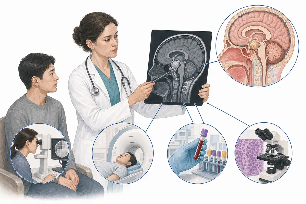
Diagnostic Criteria for Craniopharyngioma
1.Sellar region MRI (plain + contrast-enhanced)
MRI is the preferred method for diagnosing craniopharyngioma. Typical findings include a suprasellar cystic-solid mass;it shows high signal intensity on T1WI (due to cholesterol crystals) and T2WI, with associated calcification, and the cyst wall and solid components enhance after contrast administration.
2.CT bone window scan
CT is highly sensitive for detecting calcification; the presence of eggshell-like or nodular calcification in the sellar region is a characteristic feature of craniopharyngioma and serves as a key diagnostic indicator.
3.Comprehensive endocrine assessment
Measurement of hormone levels related to the thyroid, adrenal, and gonadal axes, as well as growth hormone and antidiuretic hormone, to determine the extent of pituitary-hypothalamic dysfunction and guide hormone replacement therapy.
4.Visual acuity and visual field examination
Assesses visual acuity and visual field defects; bitemporal hemianopsia is a classic sign of optic chiasm compression and serves as a key basis for determining the timing of surgery.
5.Water deprivation test
The water deprivation vasopressin test is used to diagnose diabetes insipidus and assess antidiuretic hormone secretion; it addresses a condition typically associated with hypothalamic involvement.
6.Fundus examination
Examining the optic disc for edema, pallor, or atrophy to assess the severity of long-term optic nerve compression is a vital component of the preoperative evaluation.
7.Assessment of child growth and development
Measuring height, weight, and the development of secondary sexual characteristics to assess growth hormone deficiency and the extent of delayed puberty is a core component of follow-up for pediatric patients.
8.Neuropsychological assessment
Assess cognitive function, memory, attention, and behavioral changes; hypothalamic involvement can lead to cognitive and behavioral abnormalities and serves as a key indicator for long-term follow-up.
9.Cerebrospinal fluid examination
Preoperative assessment of cerebrospinal fluid (CSF) routine and biochemical parameters is performed to rule out intracranial infection; postoperative monitoring checks for CSF leakage and intracranial infection.
Daily Self-Check
Children exhibiting unexplained vision loss, excessive thirst and urination, or short stature should seek medical attention promptly.Regular endocrine check-ups and vision assessments facilitate early detection.
Conclusion
Diagnosis relies on MRI of the sellar region (revealing cystic-solid masses and calcification) and endocrine testing.Multidisciplinary collaboration and long-term follow-up are crucial for the patient's quality of life.


