


1. Embryonic remnants
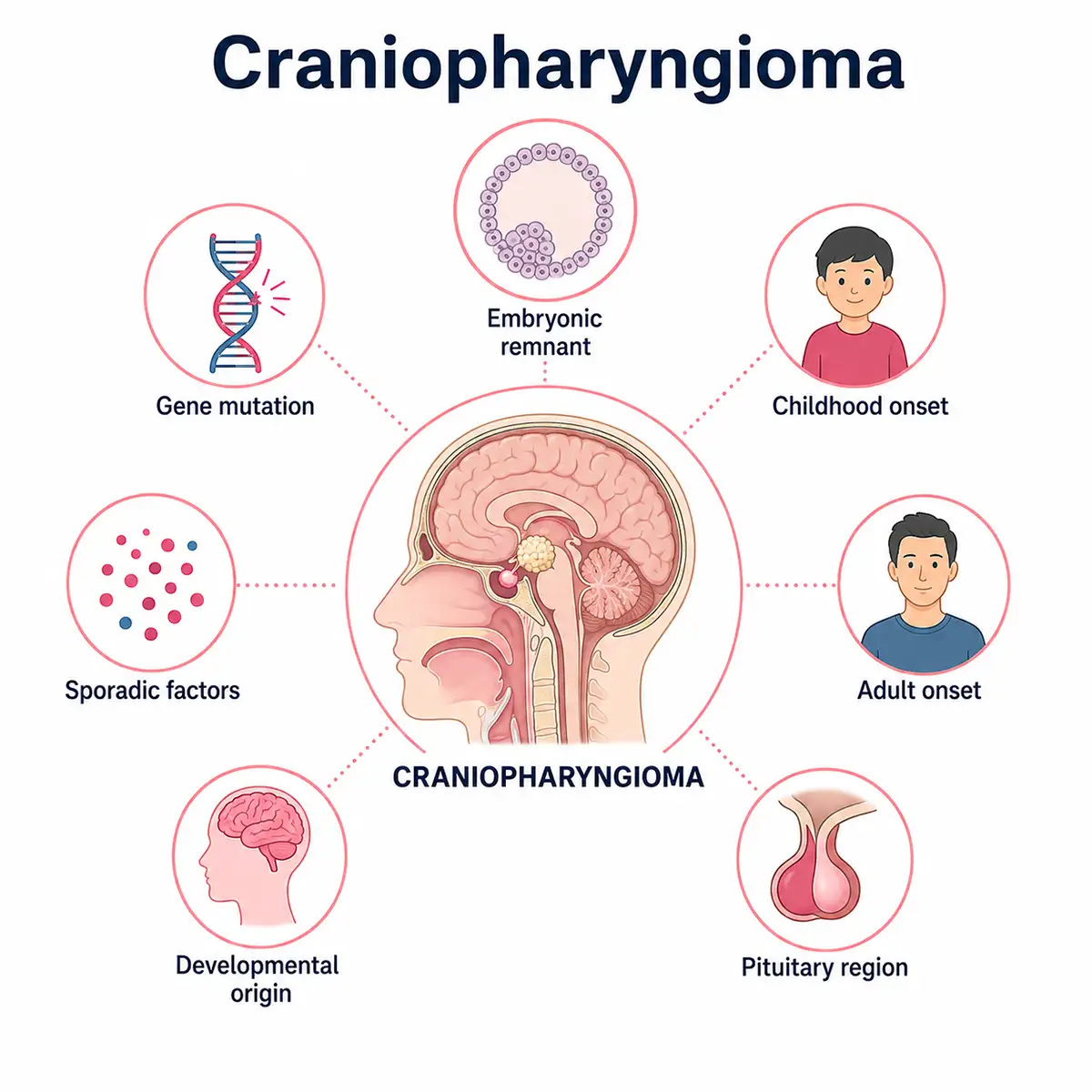
Craniopharyngiomas arise from residual epithelial tissue of the craniopharyngeal duct; incomplete regression of the duct during early pregnancy can lead to the formation of primitive cells that give rise to the tumor.
2. Rathke's cleft
Incomplete closure of Rathke's cleft during embryonic development allows residual squamous epithelial cells to proliferate in the pituitary stalk or sellar region, forming a tumor.
3. CTNNB1 mutation
Adamantinomatous craniopharyngiomas harbor *CTNNB1* gene mutations, leading to aberrant activation of the Wnt signaling pathway, which promotes tumor growth.
4. BRAF mutation
Papillary craniopharyngiomas harbor the BRAF V600E gene mutation, which leads to constitutive activation of the MAPK signaling pathway, driving tumor formation.
5. Age distribution
The adamantinomatous subtype is more common in children and adolescents, whereas the papillary subtype is more common in adults; the two subtypes exhibit distinct molecular genetic characteristics.
6. Genetic factors
A very small number of cases are associated with familial inheritance; while *CTNNB1* mutations can be inherited, the majority of cases are sporadic.
7. Gender differences
The incidence of craniopharyngioma is roughly equal between boys and girls—or slightly higher in boys—showing no significant gender predilection;it is a common pediatric tumor in the sellar region.
Conclusion
Craniopharyngioma arises from a combination of factors, including embryonic developmental abnormalities and genetic mutations. Although benign, the tumor can compress the optic nerve and pituitary gland, leading to serious consequences.Prompt medical attention is crucial if children exhibit vision loss or developmental delays, as early detection and timely intervention are vital.


