


Craniopharyngioma is a tumor resulting from the abnormal proliferation of residual tissue from the embryonic craniopharyngeal duct.The craniopharyngeal duct is a passage connecting the oral cavity and the pituitary gland during embryonic development; while it normally regresses and disappears, residual tissue can undergo neoplastic transformation.
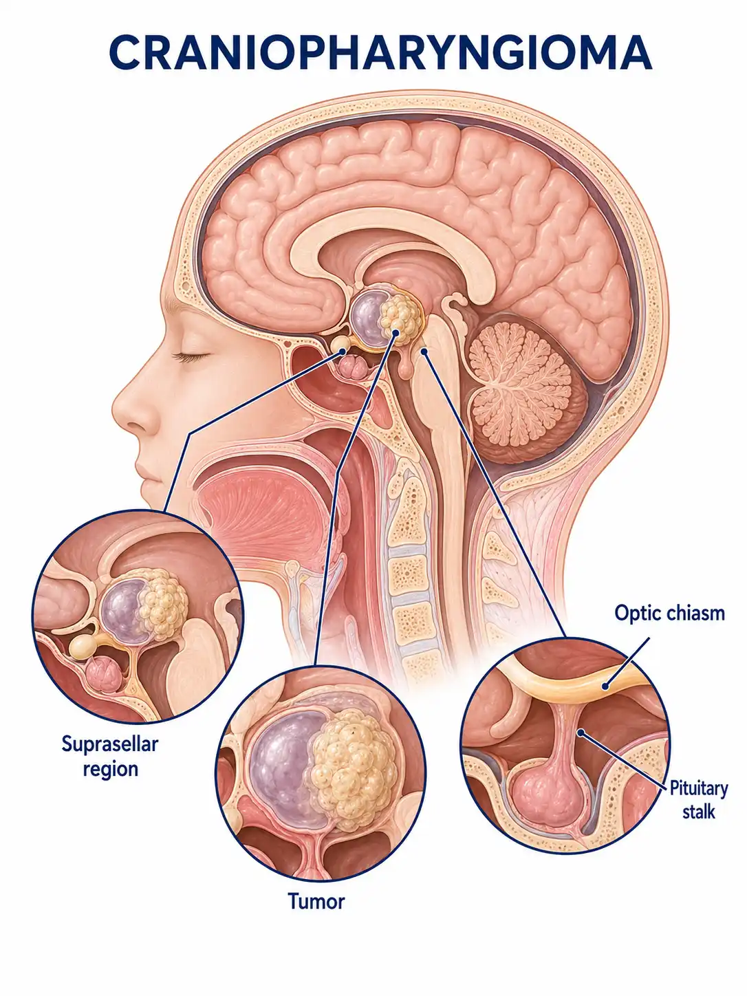
Global overview of craniopharyngioma
Craniopharyngiomas account for approximately 2%–5% of all intracranial tumors and are the most common tumors in the pediatric sellar region. They can occur at any age, with two peak incidence periods: children aged 5–14 and adults aged 50–74.
Consult a doctor regarding cancer-related issues
Children or adults presenting with symptoms such as vision loss, excessive thirst and urination, delayed growth and development, or headache should promptly consult a neurosurgeon or endocrinologist.
Main types of craniopharyngioma
1. Ameloblastomatous type
It is commonly seen in children;pathologically, it is characterized by squamous epithelial cells arranged in nests, accompanied by calcification and cystic changes, and is prone to recurrence.
2. Squamous-papillary type
It is commonly seen in adults; the cells are well-differentiated, cystic components are minimal, and the prognosis is relatively favorable.
3. Cystic craniopharyngioma
The tumor is predominantly cystic, with fluid resembling engine oil or containing cholesterol crystals; it causes symptoms by compressing surrounding structures.
4.Solid craniopharyngioma
The tumor is predominantly solid and rare; it is tightly adherent to surrounding tissues, making surgery difficult.
5. Mixed-type craniopharyngioma
It contains both solid and cystic components with frequent calcification and represents the most common form of presentation.
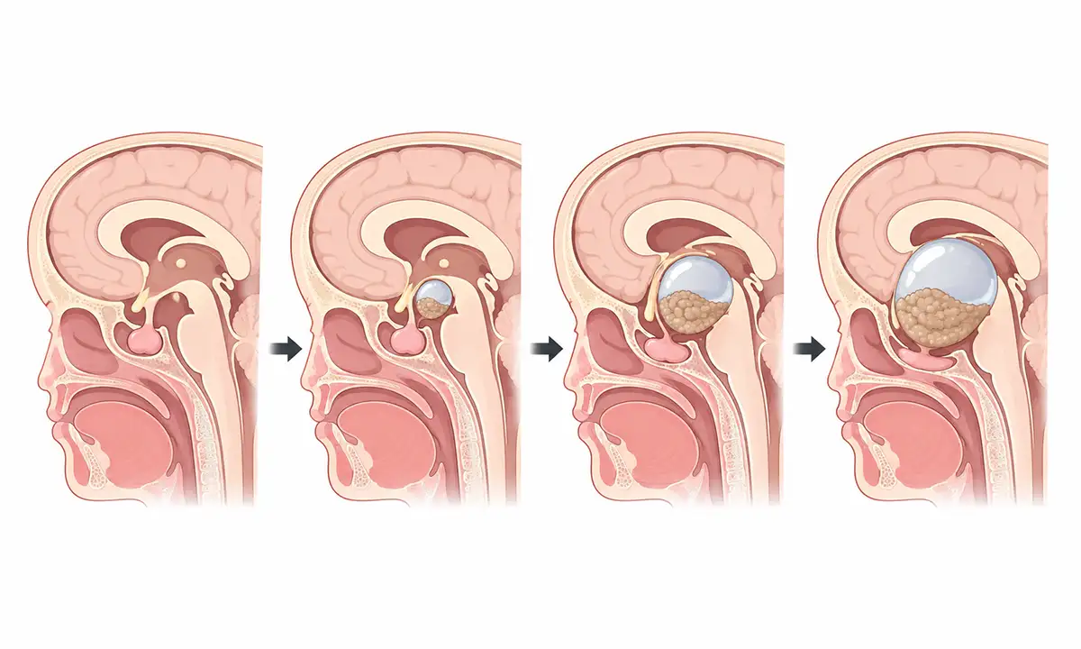
Typical symptoms and signs of craniopharyngioma
1. Visual impairment
Bitemporal hemianopia and vision loss resulting from tumor compression of the optic chiasm are the most common initial symptoms of craniopharyngiomas.
2. Excessive thirst and urination
Tumor compression or damage to the hypothalamus-pituitary stalk leads to central diabetes insipidus, manifesting as polydipsia, polyuria, and nocturia.
3. Delayed growth and development
Short stature and developmental delay caused by growth hormone deficiency are common reasons for pediatric consultations.
4. Headache
Headache occurs due to tumor compression of surrounding structures or hydrocephalus, typically located in the frontal region or retro-orbital area.
5. Endocrine disorders
Hypopituitarism leads to reduced function of the thyroid, adrenal glands, and gonads, resulting in fatigue, cold intolerance, and diminished sexual function.
6. Obesity
Hypothalamic involvement leads to impaired regulation of food intake, resulting in weight gain and obesity that are difficult to manage through dietary control.
Diagnostic Methods for Craniopharyngioma
1. Hormone level testing
Comprehensively assess pituitary hormone levels—including growth hormone, thyroid hormone, cortisol, and sex hormones—to evaluate the functional status of the pituitary gland.
2. Sellar region MRI
Contrast-enhanced MRI is the preferred method for diagnosing craniopharyngioma; typical findings include a suprasellar cystic-solid mass with calcification.
3. Cranial CT
CT can clearly visualize intratumoral calcifications and serves as an important tool for assessing the extent of calcification.
4. Visual acuity and visual field examination
Ophthalmological examinations assess visual acuity and visual field defects; bitemporal hemianopia is a typical sign of optic chiasm compression.
5. Water deprivation test
For the diagnosis and differential diagnosis of central diabetes insipidus, and for assessing the severity of antidiuretic hormone deficiency.
6. Bone age assessment
Bone age assessment is performed in pediatric patients to evaluate the severity of growth and developmental delay.
Treatment methods for craniopharyngioma
1. Surgical treatment
Tumor resection via endoscopic endonasal surgery or craniotomy is the primary treatment modality; the goal is to relieve compression while preserving hypothalamic function.
2. Cyst fluid drainage
For tumors predominantly composed of cystic components, stereotactic puncture and drainage of the cystic fluid can be performed to alleviate symptoms.
3. Radiotherapy
Postoperative adjuvant radiotherapy can reduce recurrence rates, and proton therapy can minimize damage to surrounding normal tissues.
4. Hormone replacement
Lifelong hormone replacement therapy is required postoperatively, including thyroxine, glucocorticoids, growth hormone, and sex hormones.
5. Treatment of diabetes insipidus
Desmopressin is used to control symptoms of diabetes insipidus and maintain water-electrolyte balance.
6. Chemotherapy perfusion
Intracystic chemotherapy drug perfusion can be used to treat recurrent cystic craniopharyngioma.
7. Nutritional management
Involves dietary control and exercise guidance to manage hypothalamic obesity, with metabolic intervention implemented when necessary.
8. Follow-up monitoring
Postoperative follow-up involving regular MRI of the sellar region and hormone level testing is required to monitor for tumor recurrence and endocrine status.
Conclusion
Craniopharyngioma is a common congenital tumor of the sellar region; although benign, it is deeply situated and presents treatment challenges.Surgery, radiotherapy, and standardized hormone replacement therapy help improve patient prognosis and quality of life.


