


Global disease prevalence: Chordoma is an extremely rare malignant tumor worldwide; it typically occurs at the skull base and sacrococcygeal region, is highly locally invasive, and is prone to recurrence.
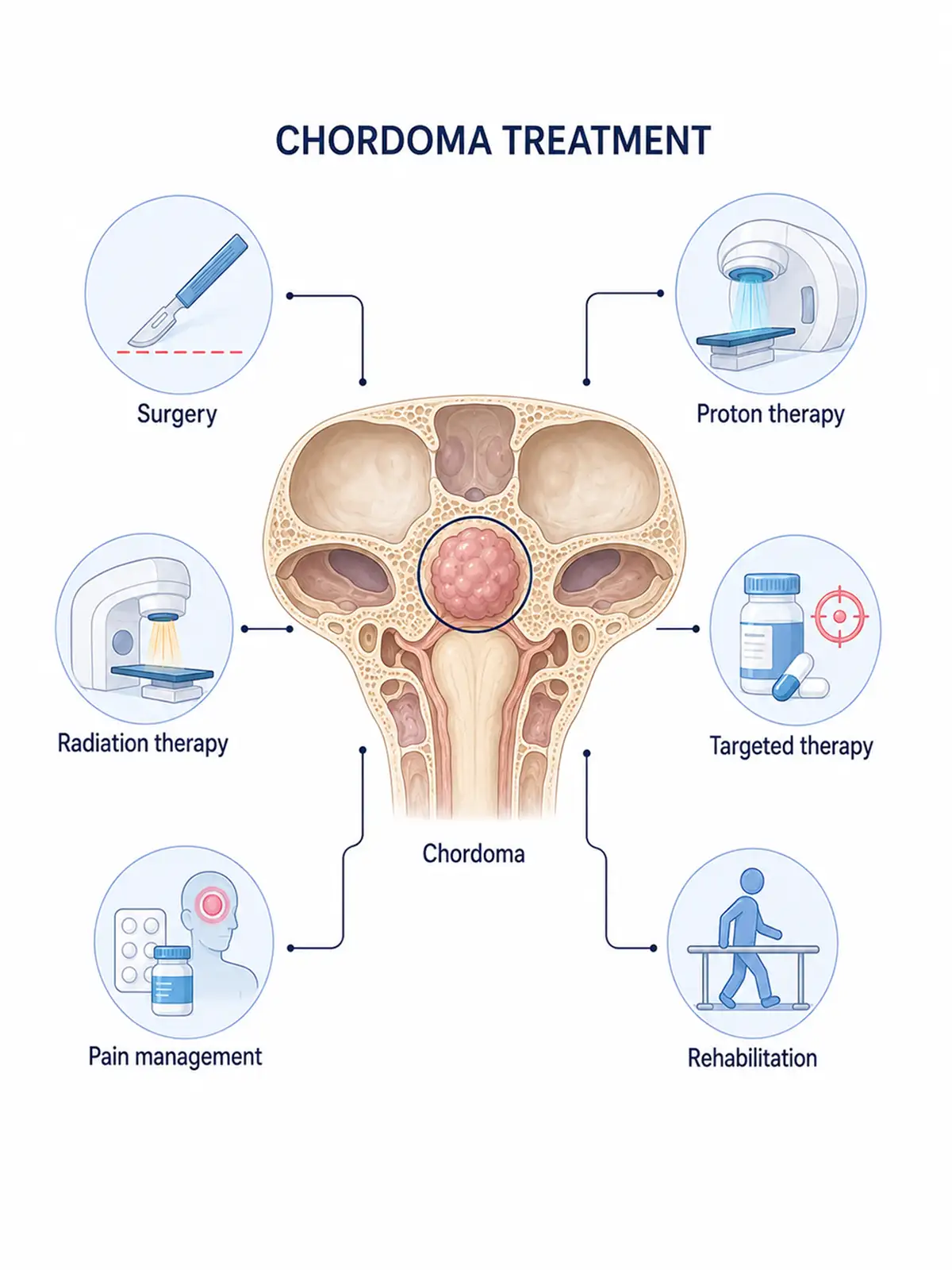
Staged treatment for chordoma
Early treatment (involving a localized, small tumor with a favorable response)
specifically surgical resection combined with adjuvant therapy—can effectively control tumor progression, and the patient's prognosis is relatively good.
Intermediate-stage treatment (addressing local spread, inhibiting growth, and delaying progression)
employs a comprehensive approach—combining surgery with radiotherapy—to control further tumor spread and slow the rate of disease deterioration.
Treatment for advanced-stage disease (involving distant metastasis, symptom relief, and survival prolongation)
primarily relies on systemic therapies such as radiotherapy and targeted therapy to alleviate clinical symptoms and extend patient survival as much as possible.
Consequences of failure to receive timely treatment in the early stage: If standardized treatment is not administered promptly in the early stage, the tumor will continue to grow and spread to surrounding bone tissue and nerves; missing the optimal window for intervention will lead to disease progression.
Why choose Fosun Health for chordoma treatment
Leveraging its hospitals' platforms—including M6 CyberKnife, VitalBeam, neurosurgery/skull base surgery, imaging, and medical physics—Fosun Health facilitates multidisciplinary consultations involving experts in neurosurgery, radiotherapy, radiology, pathology, and rehabilitation.The team formulates a treatment plan combining maximal safe resection with precision radiotherapy, taking into account tumor location, surgical margins, prior treatments, and the involvement of the brainstem, spinal cord, and cranial nerves.
Microsurgery emphasizes preserving neurological function and structural stability while resecting the tumor; CyberKnife or fractionated radiotherapy can optimize radiation dosage for postoperative residual or localized recurrent lesions, minimizing exposure to the brainstem, spinal cord, visual pathways, and normal tissues, thereby reducing the risk of certain treatment-related adverse effects.Fosun Health also provides long-term MRI or CT follow-up and neurological rehabilitation support.

Chordoma Treatment Methods
1.M6 CyberKnife treatment
Suitable for chordomas of the skull base or spine presenting with small residual tumor volumes post-surgery, localized recurrence, or inoperability, provided that radiation doses to critical organs remain within safety limits.
Treatment advantages:
① Targeting localized residual disease: Suitable for select small post-operative lesions.
② Protection of critical neural structures: Reduces radiation dose to the brainstem, spinal cord, and visual pathways.
③ Suitable for complex skull base anatomy: Enables dose shaping using multi-angle radiation beams.
④ Strict verification of previous doses: The original treatment plan must be reconstructed prior to re-irradiation for recurrence.
2.VitalBeam precision radiotherapy
Indicated for skull base and spinal chordomas that are residual post-surgery, unresectable in their entirety, large in volume, or require high-dose fractionated radiotherapy.
Treatment advantages:
① Covers the surgical bed and invaded bone: Suitable for large, irregular target areas.
② Protect brainstem and spinal cord: Control radiation dose to critical organs using intensity-modulated planning.
③ Support high-dose fractionation: Increases the local radiation dose while remaining within the tolerance limits of normal tissues.
④ Suitable for postoperative comprehensive therapy: Reduces the risk of further growth of residual lesions.
3.Resection and stabilization/reconstruction for skull base or spinal chordoma
Indicated for primary or recurrent chordomas where anatomical conditions allow, the patient can tolerate the procedure, and maximal resection is achievable.
4.Molecular targeted therapy or clinical trial
Indicated for chordomas that cannot be controlled by local therapy, or that are recurrent, progressive, or metastatic; selection is based on molecular testing and clinical conditions.
Attend regular follow-up appointments and actively cooperate with subsequent treatments.
5.Proton beam therapy
Uses proton beams to precisely target tumor tissue while maximizing protection of surrounding normal tissue; suitable for patients with skull base chordoma.Helps improve therapeutic efficacy.
6.Heavy ion therapy
Utilizes heavy-ion beams to precisely destroy tumor tissue; it is effective for treating chordomas that are resistant to conventional radiotherapy, helping to enhance therapeutic outcomes.
Post-treatment examinations
Periodic follow-up imaging and tumor marker testing are required after treatment to timely detect tumor recurrence or changes in the disease status.
Conclusion
Although rare, chordoma poses a serious threat; successful treatment hinges on early detection, diagnosis, and intervention, as well as selecting an appropriate treatment plan and adhering to follow-up care.


