


Chordoma is a very rare malignant tumor, accounting for approximately 0.15%–0.2% of all bone tumors.It typically affects individuals aged 40–70, with a slightly higher incidence in men than in women.About one-third of cases occur at the clivus (skull base), one-third in the sacrococcygeal region, and one-third in the mobile spine.It has a high rate of local recurrence and the potential for distant metastasis.
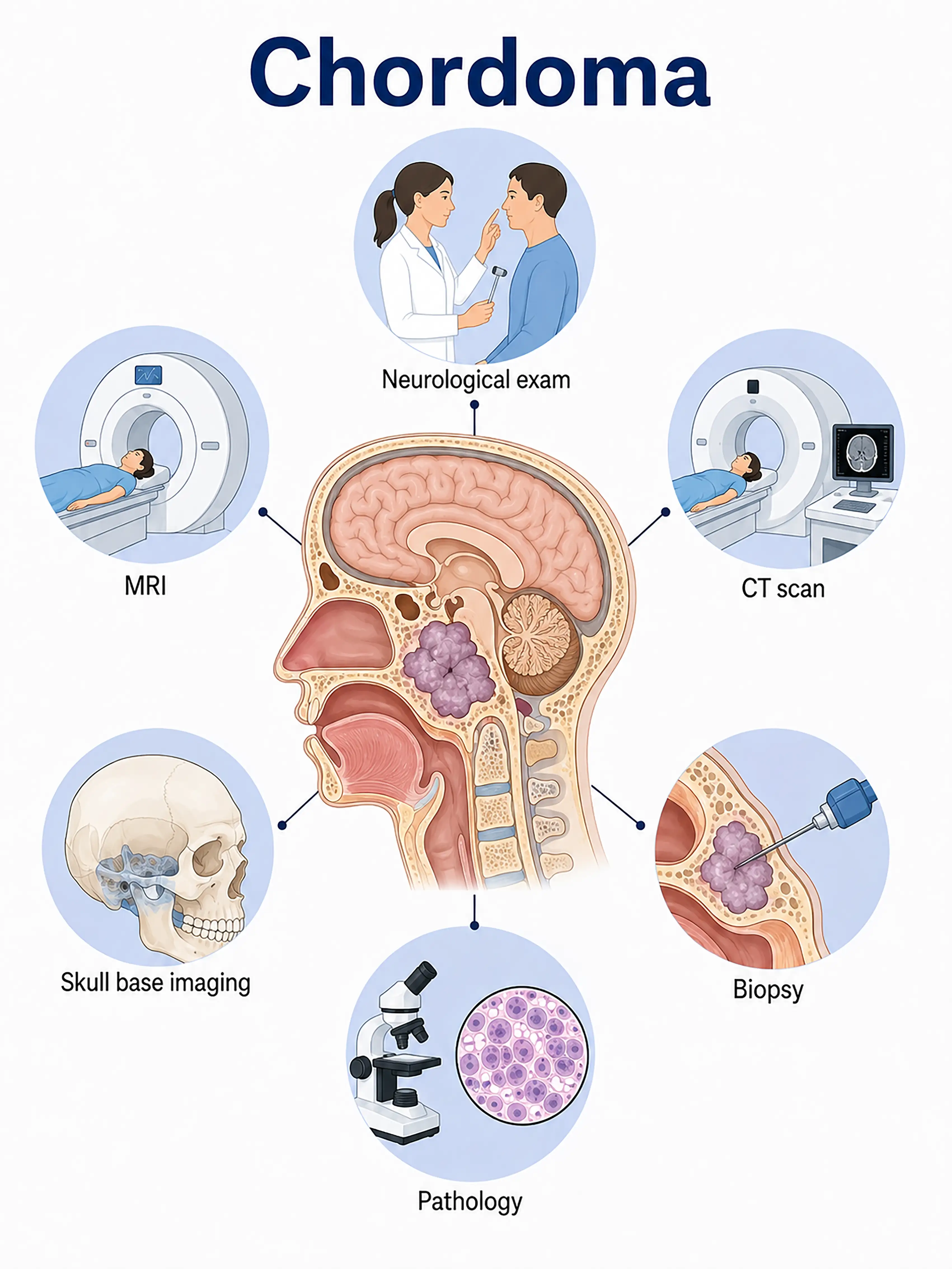
Diagnosis based on chordoma stages
1.Early diagnosis of chordoma
The tumor grows slowly, often presenting with no obvious symptoms in the early stages.MRI reveals a soft-tissue mass with hyperintensity on T2-weighted images (T2WI), while CT shows bone destruction and intratumoral calcification; biopsy is required for definitive diagnosis.
2.Diagnosis of mid-stage chordoma
Persistent headache, double vision, or lumbosacral pain occur.MRI reveals tumor enlargement with extensive bone destruction; a CT-guided needle biopsy confirms the pathological diagnosis.
3Diagnosis of advanced chordoma
Tumor extensively invades the skull base or pelvic structures; presents with impairment of multiple cranial nerve groups or dysfunction of pelvic organs.Diagnosis confirmed via surgical specimen retrieval.
If not diagnosed promptly, the tumor causes extensive bone destruction and invades surrounding vital structures, making complete surgical resection difficult and resulting in a very high rate of local recurrence.
When to Seek Medical Attention
Early signs of chordoma include persistent headache, double vision, facial numbness, and lumbosacral pain.Please consult a doctor via the "Chat with a Doctor" feature for early detection.During the online consultation, the doctor will ask about your symptoms and their duration, and guide you through the necessary imaging tests and biopsies.
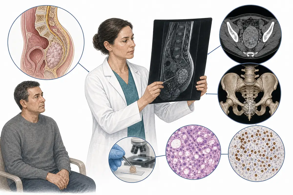
Basis for chordoma diagnosis
1.Cranial/sacrococcygeal MRI
MRI is the preferred method for diagnosing chordoma.Typical findings include a soft-tissue mass with markedly high signal intensity on T2WI and extensive bone destruction, showing heterogeneous enhancement after contrast administration.
2.CT bone window scan
CT clearly shows the extent of bone destruction and scattered intratumoral calcifications, allowing for the assessment of skull base or sacral involvement; it is a crucial tool for the preoperative evaluation of bony structures.
3.Needle biopsy pathology
CT-guided needle biopsy for pathological examination is the gold standard for diagnosing chordoma;typical pathological findings include vacuolated "signet-ring-like" cells.
4. Brachyury gene testing
Test for Brachyury (TBXT) gene expression; this serves as a specific molecular marker for chordoma and aids in differential diagnosis from other bone tumors.
5.3D reconstruction of skull base CT
3D CT reconstruction assesses the extent of skull base bone destruction and visualizes the relationship between the tumor and critical structures—such as the internal carotid artery and cavernous sinus—thereby aiding in the planning of the surgical approach.
6.Angiography assessment
Assesses the relationship between the tumor and major blood vessels (such as the internal carotid and vertebral arteries); particularly important for skull base chordomas to aid in surgical risk assessment.
7.Whole-body PET-CT assessment
Assess for distant metastasis; chordomas can metastasize to the lungs, liver, bones, and lymph nodes, allowing for the detection of metastatic lesions often missed by conventional imaging.
8.Neurological function assessment
A comprehensive evaluation of cranial nerve function (for the skull base type) or sacral nerve root function (for the sacrococcygeal type) is essential to determine the extent of nerve invasion by the tumor.
9.Intraoperative rapid frozen section pathology
Obtaining tissue during surgery for rapid frozen-section analysis allows for a preliminary assessment of the tumor's nature and guides the extent of surgical resection.
Daily Self-Check
Seek medical attention promptly if unexplained persistent headache, double vision, or lumbosacral pain occurs.If symptoms persist for years without relief, there should be a high index of suspicion for chordoma.
Conclusion
Diagnosis of chordoma relies on MRI, CT, and pathological biopsy.Brachyury gene testing aids in differential diagnosis.Surgery combined with proton therapy is the primary treatment approach.


