


Chordoma is a tumor resulting from the malignant proliferation of notochordal remnants from the embryonic stage.The notochord is the axial supporting structure during embryonic development; while it normally regresses, residual tissue can undergo neoplastic transformation.
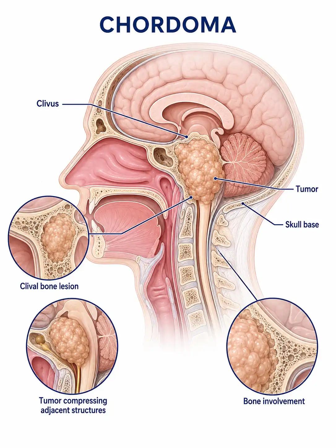
Global overview of chordoma
Chordomas account for approximately 1%–4% of all malignant bone tumors;they can occur at any age but are most common between the ages of 40 and 70. The skull base and the sacrococcygeal region are the two most common sites of occurrence.
Consult a doctor regarding cancer-related issues
Individuals experiencing symptoms such as persistent headache, double vision, sacrococcygeal pain, or difficulty with bowel or bladder function should promptly consult a neurosurgeon or an orthopedic oncologist.
Main types of chordoma
1. Conventional chordoma
The most common type; cells are arranged in cords or nests within an abundant myxoid stroma; it grows slowly but exhibits local invasiveness.
2. Chondroid chordoma
The tumor contains areas of chondroid differentiation; the prognosis is slightly better than that of the conventional type, and it requires differentiation from chondrosarcoma.
3. Dedifferentiated chordoma
Contains high-grade sarcomatous components; highly malignant, rapidly growing, prone to distant metastasis, and associated with an extremely poor prognosis.
4. Skull base chordoma
Occurs in the clivus region of the skull base, compressing the brainstem and cranial nerves; surgically challenging, representing the most difficult-to-treat type of chordoma.
5. Sacrococcygeal chordoma
Occurs in the sacrococcygeal region; can grow to a large size, compressing the rectum and bladder; surgical planning must consider the preservation of bowel and bladder function.
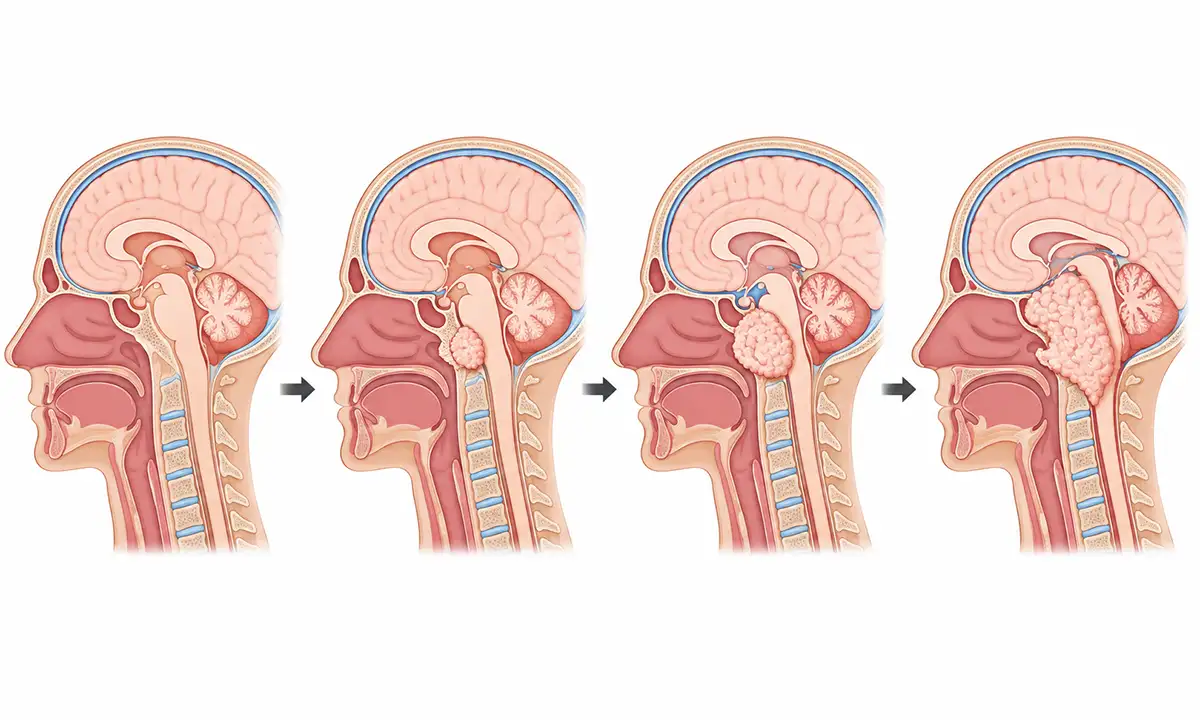
Typical symptoms and signs of chordoma
1. Headache
Compression of the brainstem and cranial nerves by a skull base chordoma causes persistent headache—typically located in the occipital region—accompanied by neck stiffness.
2. Double vision (diplopia)
Tumor compression of the abducens and oculomotor nerves causes diplopia and impaired eye movement.
3. Facial numbness
Facial sensory loss and pain occur when the tumor compresses the trigeminal nerve.
4. Sacrococcygeal pain
Sacrococcygeal chordoma causes persistent pain in the sacral region; the pain worsens when sitting and is notably pronounced at night.
5. Difficulty with defecation and urination
Compression of the rectum and bladder by a sacrococcygeal tumor results in constipation, difficulty defecating, and urinary frequency and urgency.
6. Lower limb weakness
Compression of sacral nerve roots by the tumor results in lower-limb weakness, numbness, and paresthesia.
Diagnostic methods for chordoma
1. Imaging examination
MRI and CT visualize tumor location, size, and bone destruction; typical findings include osteolytic destruction accompanied by a soft-tissue mass.
2. Pathological biopsy
Pathological examination of tumor tissue obtained via needle biopsy or surgery typically reveals cords of cells floating in a myxoid matrix.
3. Immunohistochemistry
Positivity for S-100 protein, EMA, and brachyury aids in the diagnosis and differential diagnosis of chordoma.
4. Molecular testing
Detection of mutations in genes associated with the PI3K/AKT/mTOR pathway provides a basis for targeted therapy.
5. Systemic assessment
Whole-body bone scanning and PET-CT are used to assess for distant metastases; the lungs are the most common site of metastasis.
6. Neurological function assessment
A detailed assessment of cranial and sacral nerve function provides a basis for formulating the surgical plan.
Chordoma treatment methods
1. Surgical treatment
Wide resection is the primary treatment for chordoma; however, due to the tumor's unique location, achieving ideal surgical margins is often difficult.
2. Proton therapy
Proton therapy delivers high-dose radiation to the tumor while sparing surrounding critical structures, serving as an effective adjuvant treatment for chordoma.
3. Radiotherapy
Intensity-modulated radiotherapy (IMRT) and stereotactic radiotherapy can be used as adjuvant therapy after surgery or for cases where surgery is not feasible.
4. Targeted therapy
Targeted drugs addressing the EGFR and mTOR pathways are under investigation, offering new treatment options for relapsed or refractory chordoma.
5. Interventional embolization
Preoperative embolization reduces tumor blood supply and lowers the risk of intraoperative hemorrhage.
6. Pain management
Effectively manage pain in patients with advanced disease to improve quality of life.
7. Functional rehabilitation
Postoperative neurological rehabilitation training improves motor and sensory functions.
8. Follow-up monitoring
Long-term, regular postoperative MRI follow-up is necessary to monitor for local recurrence; chordomas have a high recurrence rate, requiring lifelong follow-up.
Conclusion
Chordoma is a locally aggressive malignant tumor originating from notochordal remnants, commonly occurring at the skull base and in the sacrococcygeal region.Comprehensive treatment approaches, such as wide resection combined with proton therapy, help control tumor progression.


