


Astrocytoma is a tumor formed by the abnormal proliferation of astrocytes.Astrocytes are the most abundant type of glial cell in the central nervous system, performing functions such as supporting neurons, maintaining the blood-brain barrier, and repairing damage.
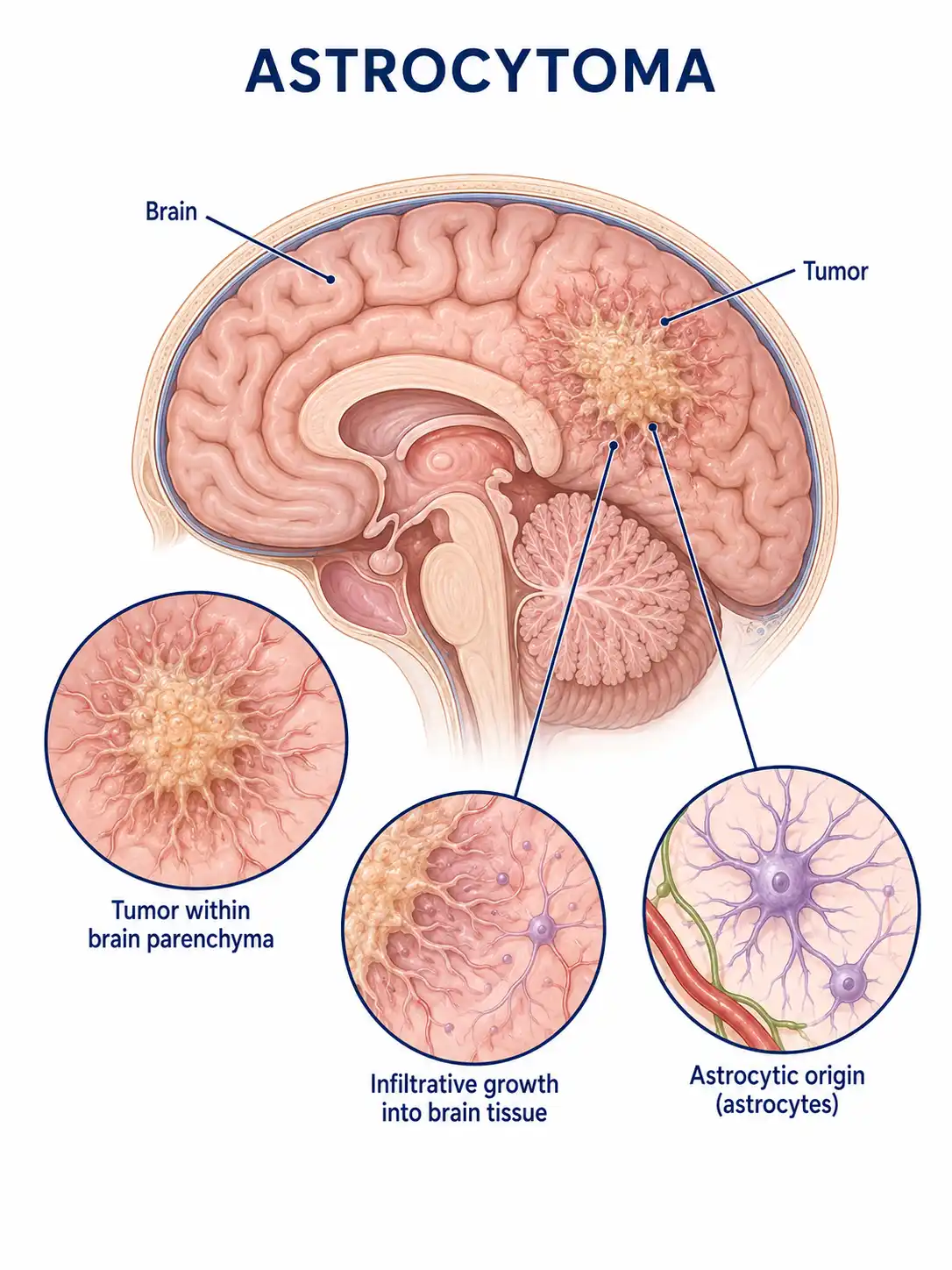
Global overview of astrocytoma
Astrocytomas account for a significant proportion of gliomas and can occur at any age; certain low-grade types are more common in children.High-grade astrocytomas have a poor prognosis and are a key focus of neuro-oncology research.
Consult a doctor regarding cancer-related issues
Individuals experiencing symptoms such as new-onset seizures, persistent headache, limb weakness, or cognitive decline should promptly consult a neurosurgeon or a neuro-oncologist for evaluation.
Major types of astrocytoma
1. Pilocytic astrocytoma
WHO Grade I;slow-growing with well-defined margins; common in children and adolescents; typically arises in the cerebellum and optic nerve; prognosis is favorable following complete surgical resection.
2. Diffuse astrocytoma
WHO Grade II; slow-growing but infiltrative; difficult to completely resect; carries a risk of progression to a high-grade tumor.
3. Anaplastic astrocytoma
WHO Grade III;characterized by relatively rapid growth and marked cellular atypia; requires comprehensive treatment combining surgery with radiotherapy and chemotherapy.
4. Glioblastoma
WHO Grade IV; the most malignant form of astrocytoma; rapid growth; highly aggressive; prone to recurrence; extremely poor prognosis.
5. Pleomorphic xanthoastrocytoma
A relatively rare type; it commonly affects children and adolescents, is often located in the superficial regions of the cerebral hemispheres, and is mostly low-grade.
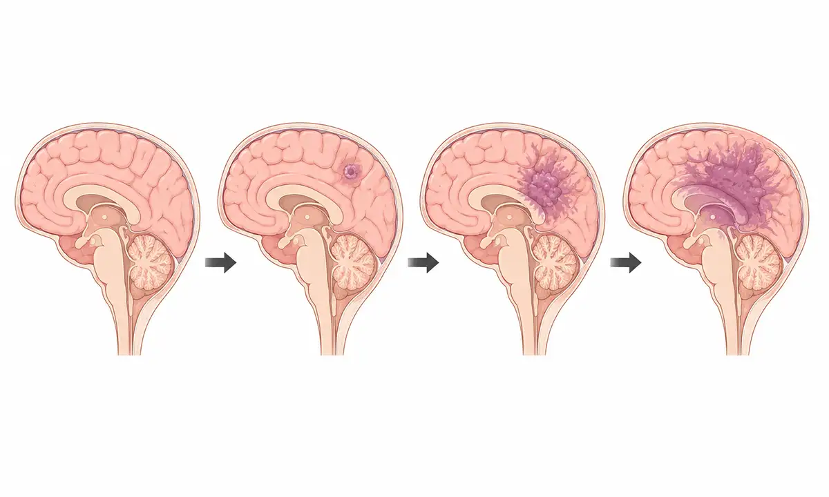
Typical symptoms and signs of astrocytoma
1. Epileptic seizure
New-onset epilepsy is a common initial symptom of astrocytomas, particularly low-grade tumors, and may manifest as focal or generalized seizures.
2. Headache and nausea
A headache that gradually worsens—particularly in the morning—and is accompanied by nausea and vomiting is a classic sign of increased intracranial pressure.
3. Limb weakness
Compression of the motor functional area or conduction tracts by the tumor results in weakness, numbness, or poor coordination in the limbs on one side.
4. Speech difficulty
When the tumor involves speech-related functional areas, difficulties in speech expression or comprehension—including word-finding difficulties—arise, affecting daily communication.
5. Changes in vision
Vision loss, visual field defects, or diplopia occur when a tumor compresses the visual pathways or causes increased intracranial pressure.
6. Cognitive decline
Frontal or temporal lobe tumors can cause memory loss, inattention, personality changes, and impaired judgment.
Diagnostic Methods for Astrocytoma
1. Neurological examination
Detailed examinations include assessments of visual acuity, muscle strength, sensation, reflexes, and coordination to evaluate the extent of neurological deficits and localize the lesion.
2. Cranial MRI scan
MRI is the preferred method for diagnosing astrocytoma; it reveals tumor location, size, and surrounding edema, while contrast-enhanced scanning aids in determining the tumor grade.
3. Pathological biopsy
Obtaining tumor tissue via surgical resection or needle biopsy for pathological examination is the gold standard for diagnosis and grading.
4. Molecular testing
Detection of molecular markers such as IDH mutations, MGMT methylation, and TERT promoter mutations provides valuable guidance for prognostic assessment and treatment selection.
5. Functional MRI
Functional MRI can localize language and motor functional areas, aiding in surgical planning and the preservation of critical brain regions.
6. Spectroscopic analysis
Magnetic resonance spectroscopy (MRS) can detect the metabolic characteristics of tumor tissue, aiding in the differentiation of tumor grades and the distinction between tumor recurrence and radiation necrosis.
What are the treatment methods for astrocytoma
1. Surgical treatment
Surgery is the primary treatment for astrocytoma; the goal for low-grade tumors is gross total resection, while high-grade tumors undergo maximal safe resection.
2. Radiation therapy
Postoperative adjuvant radiotherapy is indicated for high-grade astrocytomas to reduce the risk of local recurrence; proton therapy can minimize damage to normal brain tissue.
3. Chemotherapy
Temozolomide is the standard chemotherapy drug for high-grade astrocytomas, whereas the PCV regimen is used for tumors with a predominant oligodendroglial component.
4. Targeted therapy
Targeted drugs addressing the BRAF V600E mutation can be used for specific types of astrocytoma, while bevacizumab can alleviate symptoms.
5. Electric field therapy
Tumor-treating fields (TTFields) combined with chemoradiotherapy can be used for newly diagnosed glioblastoma; they inhibit tumor growth by interfering with cell division.
6. Immunotherapy
Immune checkpoint inhibitors and CAR-T cell therapies are currently being actively explored and researched for recurrent glioma.
7. Symptomatic and supportive care
Antiepileptic drugs control seizures, glucocorticoids reduce cerebral edema, and rehabilitation therapy addresses neurological deficits.
8. Follow-up monitoring
Regular postoperative MRI follow-up monitors for tumor recurrence and progression, with individualized follow-up plans formulated based on molecular characteristics.
Conclusion
Astrocytomas are the most common type of brain glioma, exhibiting significant differences in biological behavior between low-grade and high-grade forms.A comprehensive approach combining surgery, radiotherapy, chemotherapy, and novel therapies helps improve patient prognosis and quality of life.


