


Astrocytoma is the most common type of glioma.The 2016 WHO classification incorporated IDH mutation status into diagnostic criteria, as IDH-mutant and IDH-wildtype tumors differ significantly in prognosis and response to treatment.Grade IV astrocytoma (glioblastoma) is the most common and most malignant type of adult brain tumor.
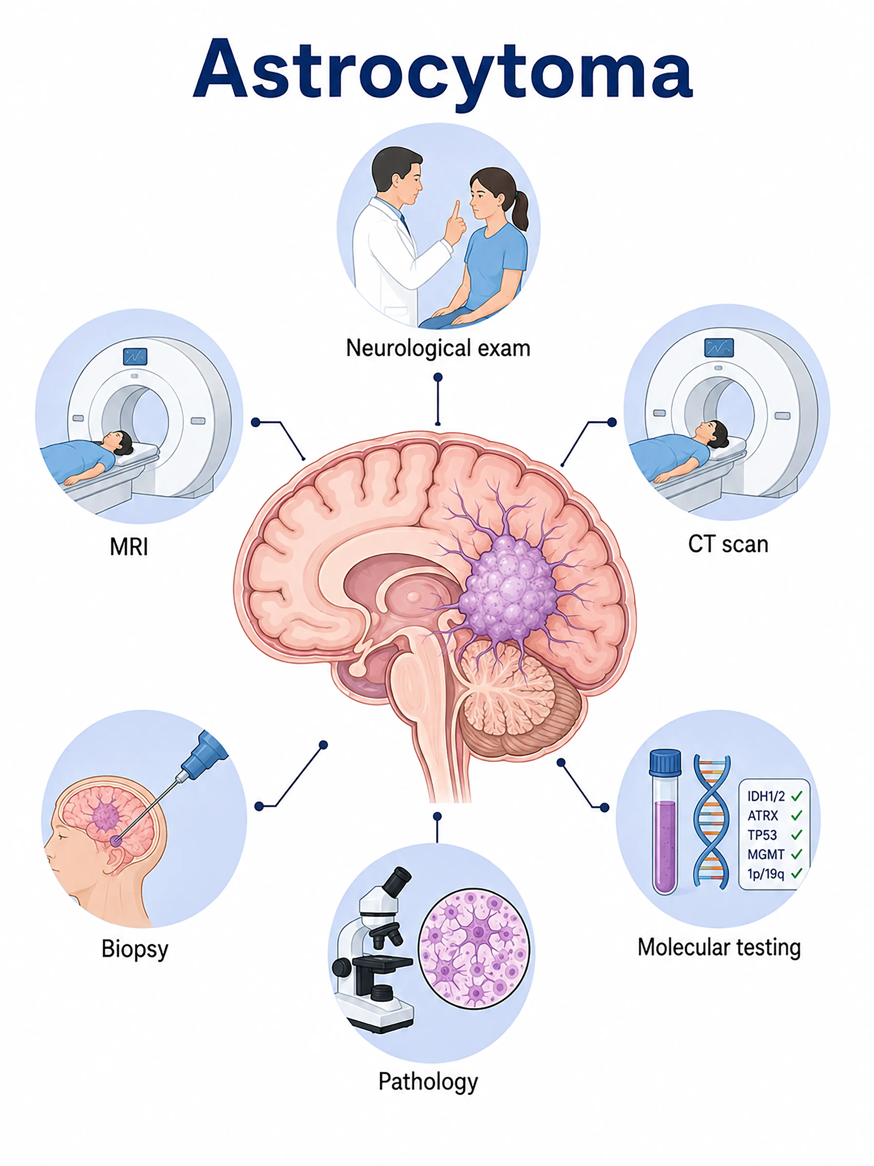
Diagnosis based on astrocytoma stage
1.Early diagnosis of astrocytoma
Low-grade tumors may not cause symptoms until they have grown quite large.MRI reveals ill-defined lesions with high signal intensity on T2/FLAIR sequences; there is no significant or only mild enhancement following contrast administration, necessitating a biopsy for definitive diagnosis.
2.Intermediate-stage astrocytoma diagnosis
Worsening headache, seizures, or neurological deficits.MRI shows lesion enlargement accompanied by enhancement and perilesional edema.Biopsy confirms pathological grade and IDH mutation status.
3.Advanced astrocytoma diagnosis
High-grade astrocytomas (Grades III–IV) progress rapidly, causing severe intracranial hypertension and extensive neurological impairment.MRI reveals heterogeneous enhancement, necrosis, and mass effect.
If not diagnosed promptly, astrocytomas—especially high-grade tumors—progress rapidly and infiltrate brain tissue extensively, resulting in poor treatment outcomes and short survival times.
When to Seek Medical Attention
Early signs of astrocytoma include new-onset seizures, persistent headaches, speech difficulties, and limb weakness.Please consult a doctor via the "Chat with a Doctor" feature for early detection.During the online consultation, the doctor will ask about your symptoms and disease progression, and guide you through the process of obtaining a cranial MRI.
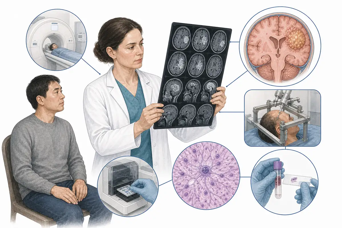
Diagnostic Criteria for Astrocytoma
1.Cranial MRI examination
MRI is the preferred imaging modality for diagnosing astrocytomas.Low-grade tumors appear hyperintense on T2/FLAIR sequences, while high-grade tumors present with heterogeneous enhancement, necrosis, and peritumoral edema.Multimodal MRI assists in grading the tumor.
2.Stereotactic biopsy
For tumors located in deep-seated areas or functional regions, stereotactic biopsy is used to obtain tissue samples for pathological examination, thereby clarifying the histological type and WHO grade.
3.Craniotomy for resection
Craniotomy and resection are performed for resectable tumors to obtain intact specimens for pathological and molecular testing while simultaneously reducing tumor burden.
4.IDH gene test
Determining IDH1/IDH2 mutation status is a key indicator for the classification of astrocytomas.IDH-mutant tumors have a more favorable prognosis, whereas wild-type tumors progress more rapidly.
5.ATRX mutation testing
ATRX mutations often coexist with IDH mutations, aiding in the differentiation between diffuse astrocytoma and oligodendroglioma.
6.MGMT methylation testing
Detection of MGMT promoter methylation status guides the selection of temozolomide chemotherapy regimens; patients with positive methylation status typically show better chemotherapy outcomes.
7.CT scan
CT can reveal calcification, hemorrhage, and bony changes, enabling rapid assessment of intracranial lesions in emergency settings; it is superior to MRI for detecting calcification.
8.PET-CT assessment
Assesses tumor metabolic activity and malignancy; aids in stereotactic biopsy localization and the differentiation between tumor recurrence and radiation necrosis.
9.Intraoperative MRI
Real-time intraoperative MRI guidance ensures the safest possible tumor resection, improving surgical precision and the rate of complete removal.
Daily Self-Check
Seek medical attention promptly if unexplained headache, seizures, or personality changes occur.Individuals with hereditary conditions such as neurofibromatosis should undergo regular neurological examinations.
Conclusion
Diagnosis of astrocytoma relies on MRI and pathological biopsy. Molecular testing—such as for IDH mutations—is central to modern classification and is vital for prognostic assessment and treatment decision-making.


