


Thyroid cancer is the most common endocrine malignancy, with rising incidence in recent years. Women are affected about three times as often as men, usually at ages 30-50. Papillary carcinoma accounts for 80%-90% and has a favorable prognosis.
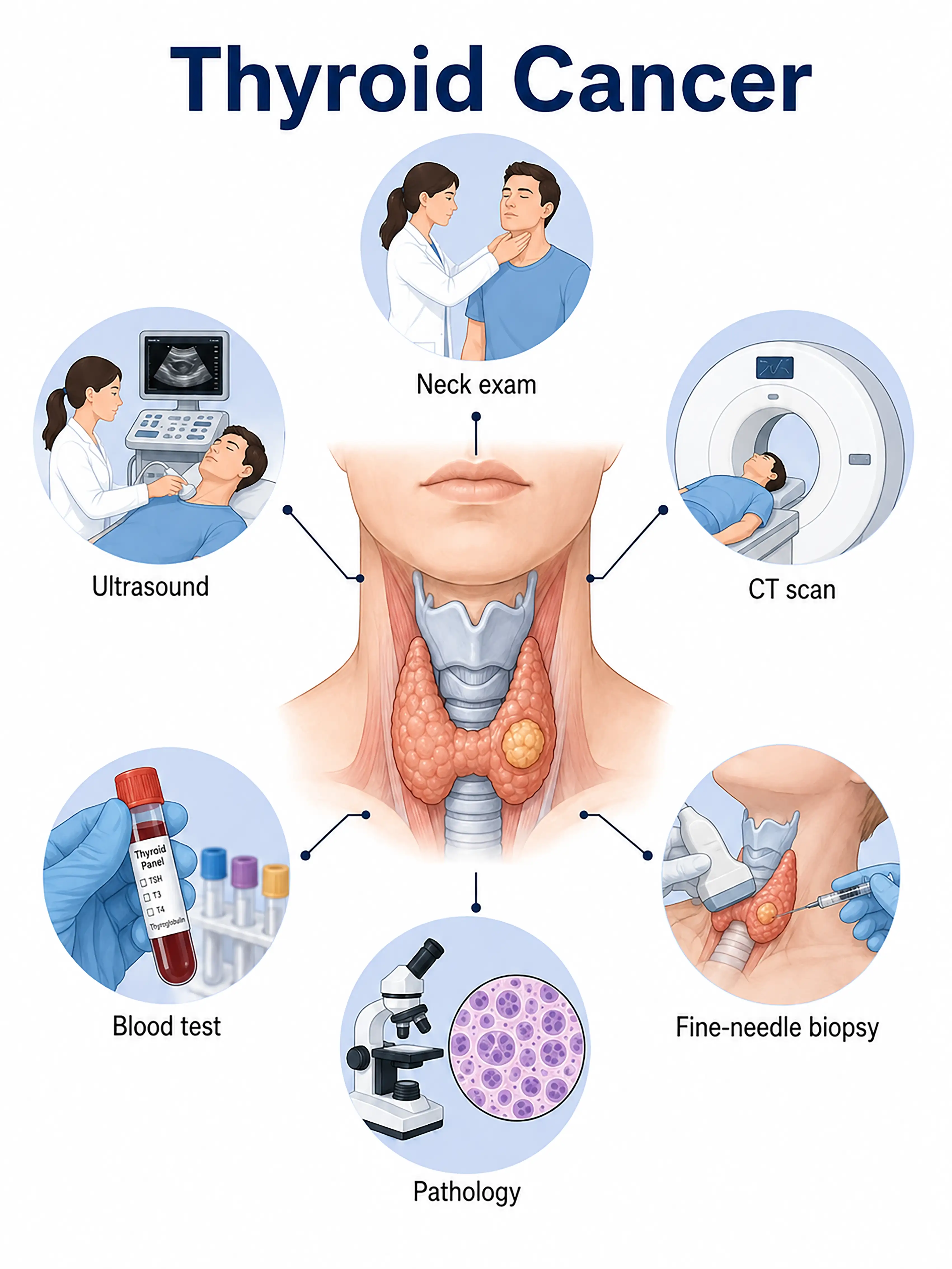
Thyroid cancer diagnosis by stage
1.Early-stage thyroid cancer diagnosis
Palpation reveals a solitary hard, poorly mobile thyroid nodule. Ultrasound shows indistinct borders, irregular shape, and microcalcifications, classified as TI-RADS category 4 or above. Fine-needle aspiration biopsy (FNA) confirms the diagnosis.
2.Intermediate-stage thyroid cancer diagnosis
The nodule enlarges, with enlarged cervical lymph nodes. Ultrasound-guided aspiration of the nodule and suspicious nodes provides cytology to establish benign or malignant status and pathological type.
3.Advanced-stage thyroid cancer diagnosis
The tumor invades the trachea, esophagus, or recurrent laryngeal nerve, or spreads to the lungs and bones. Contrast-enhanced CT and MRI assess local invasion, while whole-body PET-CT assesses distant metastasis.
Without timely diagnosis, the minority of highly malignant thyroid cancers, such as anaplastic carcinoma, rapidly invade surrounding tissues and metastasize, eliminating surgical options and resulting in an extremely poor prognosis.
When to Seek Medical Attention
Early signs of thyroid cancer include a painless neck lump, hoarseness, difficulty swallowing, and enlarged cervical lymph nodes. Consult a doctor through "Chat with a Doctor" for early detection. During an online consultation, the doctor will ask about your symptoms and radiation exposure history and guide you toward thyroid ultrasound.
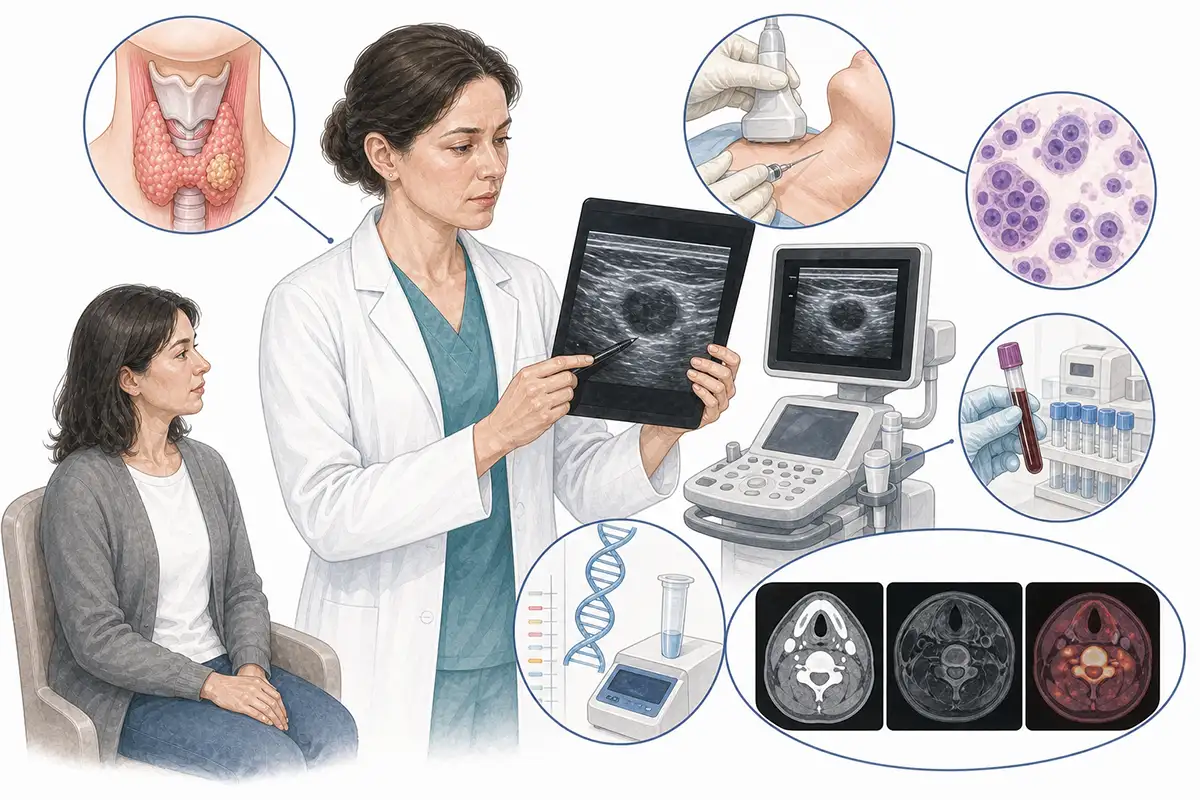
Basis for thyroid cancer diagnosis
1.Thyroid palpation
The doctor palpates thyroid size, consistency, and nodules, assessing nodule mobility, borders, and relationships to surrounding tissue. This is important for initial screening.
2.Thyroid ultrasound
Ultrasound is the preferred imaging method, assessing nodule size, shape, borders, echoes, calcification, and blood flow. TI-RADS classification guides biopsy decisions.
3.Fine-needle aspiration biopsy
Ultrasound-guided fine-needle aspiration obtains cytological samples from thyroid nodules and is the most reliable preoperative benign or malignant assessment, with accuracy exceeding 90%.
4.Thyroid function testing
Serum TSH, T3, T4, and thyroglobulin (Tg) are measured to assess thyroid function. Tg serves as a postoperative tumor marker.
5.Neck CT scan
Contrast-enhanced CT assesses invasion of the trachea, esophagus, and vessels and mediastinal and cervical lymph node metastasis, particularly important for retrosternal goiter.
6.Neck MRI
MRI assesses relationships to surrounding soft tissue and shows recurrent lesions and metastatic nodes. It is an alternative for people allergic to iodinated contrast.
7.Radionuclide scan
Iodine-131 or technetium-99m scanning assesses thyroid nodule function as hot, warm, or cold nodules to support differential diagnosis.
8.Whole-body PET-CT assessment
Whole-body PET-CT assesses distant metastasis and is important for staging radioactive iodine-refractory differentiated thyroid cancer and anaplastic carcinoma.
9.Genetic testing analysis
Testing for BRAF V600E, RAS, RET/PTC, and other genetic alterations supports diagnosis, prognosis assessment, and targeted therapy selection.
Daily Self-Check
Regularly feel the neck for thyroid lumps. Seek prompt care for growing or hardening neck nodules or hoarseness.
Conclusion
Most thyroid cancer types have a good prognosis. Ultrasound with fine-needle aspiration is central to diagnosis. Early detection and treatment can achieve long-term survival.


