


Pituitary tumors account for approximately 10%–15% of intracranial tumors.They are classified into two categories: functional (hormone-secreting) and non-functional.Functional pituitary tumors can lead to endocrine disorders such as acromegaly, Cushing's disease, and prolactinomas.Prolactinomas are the most common type in women.
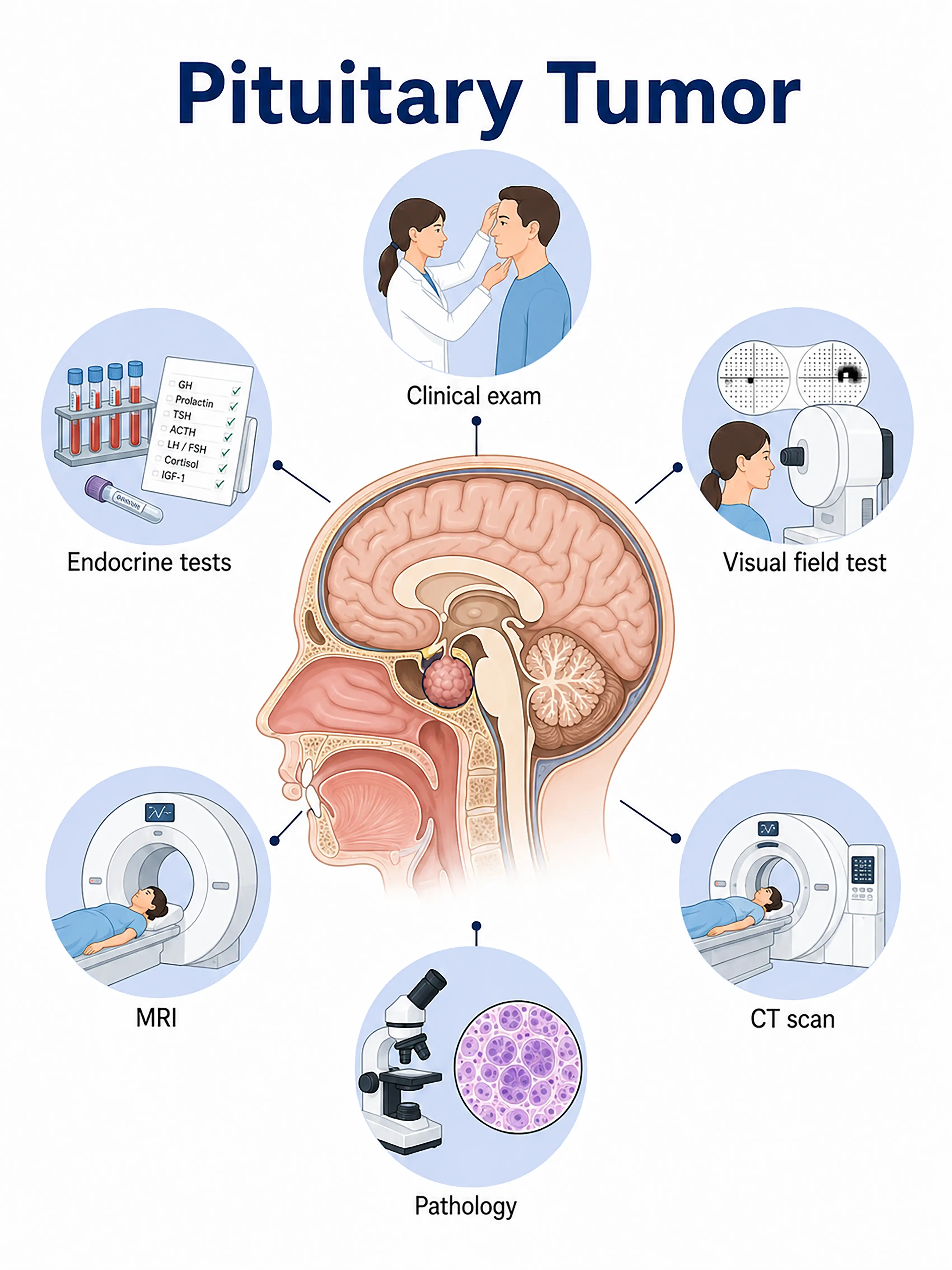
Diagnosis of pituitary tumor stages
1.Early diagnosis of pituitary tumor
Microadenomas (<1 cm) are often asymptomatic or detected due to endocrine abnormalities.Findings include abnormal serum hormone levels (PRL, GH, IGF-1, ACTH, cortisol, TSH) and a small intrasellar lesion visible on sellar MRI.
2.Diagnosis of intermediate-stage pituitary tumors
Large adenoma (≥1 cm) presenting with changes in vision/visual fields and headaches.MRI shows the tumor extending into the suprasellar region and compressing the optic chiasm; endocrine testing identifies the type of hormone secretion.
3Diagnosis of advanced pituitary tumor
A giant adenoma is invading the cavernous sinus or hypothalamus, resulting in multiple cranial nerve deficits and severe endocrine dysfunction.MRI shows extensive invasion; surgical decompression and hormone replacement therapy are required.
If pituitary tumors are not diagnosed promptly, tumor enlargement compressing the optic chiasm can result in permanent visual impairment, while long-term hormonal abnormalities associated with functional tumors can lead to severe systemic complications.
When to Seek Medical Attention
Early signs of pituitary tumors include blurred vision, visual field defects, amenorrhea, galactorrhea, and signs of acromegaly.Please consult a doctor via "Chat with a Doctor" for early detection.During the online consultation, the doctor will ask about your symptoms and endocrine changes, and guide you through hormone testing and MRI scans.
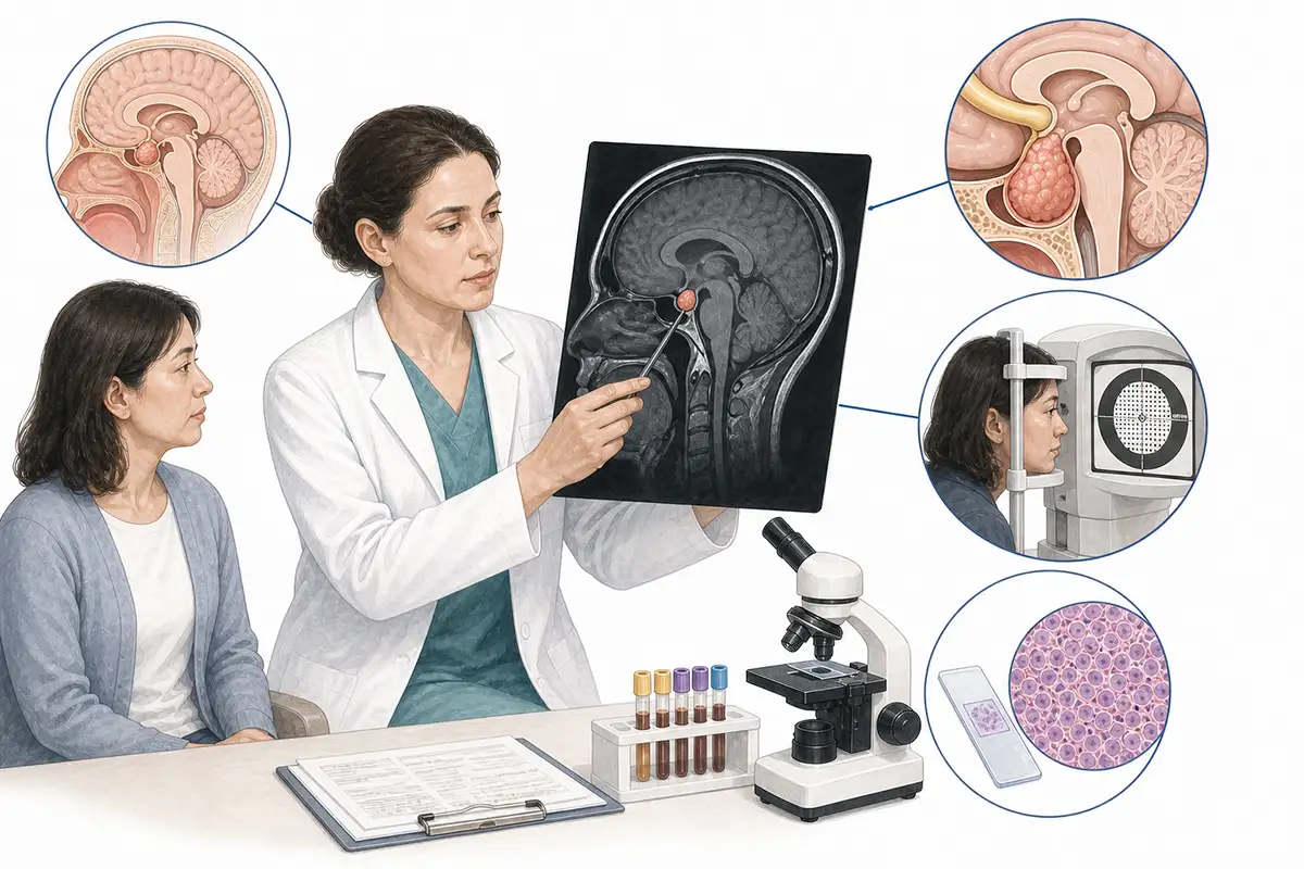
Basis for diagnosing pituitary tumors
1.Sellar region MRI (plain + contrast-enhanced)
MRI is the preferred imaging modality for diagnosing pituitary tumors.It clearly displays tumor size, location, and relationships with the optic chiasm and cavernous sinus;dynamic contrast-enhanced scanning can improve the detection rate of microadenomas.
2.Endocrine hormone testing
Measure serum levels of hormones such as prolactin (PRL), growth hormone (GH), IGF-1, ACTH, cortisol, TSH, LH, and FSH to determine the functional type of the tumor and the status of pituitary function.
3.Visual acuity and visual field examination
Standard ophthalmic examinations assess visual acuity and visual fields; bitemporal hemianopia is a classic sign of optic chiasm compression and serves as a key reference for surgical indications.
4.Fundus examination
Examining the optic disc for edema, atrophy, or color changes to assess the severity of long-term optic nerve compression is a vital component of the preoperative evaluation.
5.OGTT-GH suppression test
The oral glucose tolerance test (OGTT) is used to assess GH suppression; failure to suppress GH levels to <1 ng/mL is the gold standard for diagnosing acromegaly.
6.High-dose dexamethasone suppression test
For determining the etiology of ACTH-dependent Cushing's disease; in cases caused by pituitary tumors, high-dose dexamethasone can suppress cortisol secretion.
7.CT bone window scan
Assesses bony changes and the extent of enlargement in the sella turcica; visualizes bone resorption or destruction at the sellar floor to assist in selecting the surgical approach.
8.Comprehensive pituitary function assessment
Assessment of the functional status of the thyroid, adrenal, gonadal, and growth hormone axes to determine the presence of hypopituitarism and guide pre- and post-operative hormone replacement therapy.
9.Cavernous sinus MRI assessment
Coronal and sagittal MRI scans are used to assess the relationship between the tumor and the cavernous sinus; the Knosp grading system evaluates the extent of cavernous sinus invasion and guides the surgical plan.
Daily Self-Check
Seek medical attention promptly if unexplained changes in vision, amenorrhea with galactorrhea, or enlargement of the hands and feet occur.Regular endocrine examinations and vision assessments facilitate early detection.
Conclusion
Diagnosis of pituitary tumors relies on endocrine testing and MRI of the sellar region.Assessment of visual acuity and visual fields is a crucial basis for determining the timing of surgery, and a multidisciplinary approach represents the optimal model for diagnosis and treatment.


