


Parotid gland cancer is the most common salivary gland malignancy, accounting for about 80% of salivary gland malignancies. It usually occurs at ages 40-70, with similar incidence in men and women. Most parotid tumors are benign, with about 15%-20% malignant. Mucoepidermoid and adenoid cystic carcinomas are the most common pathological types.
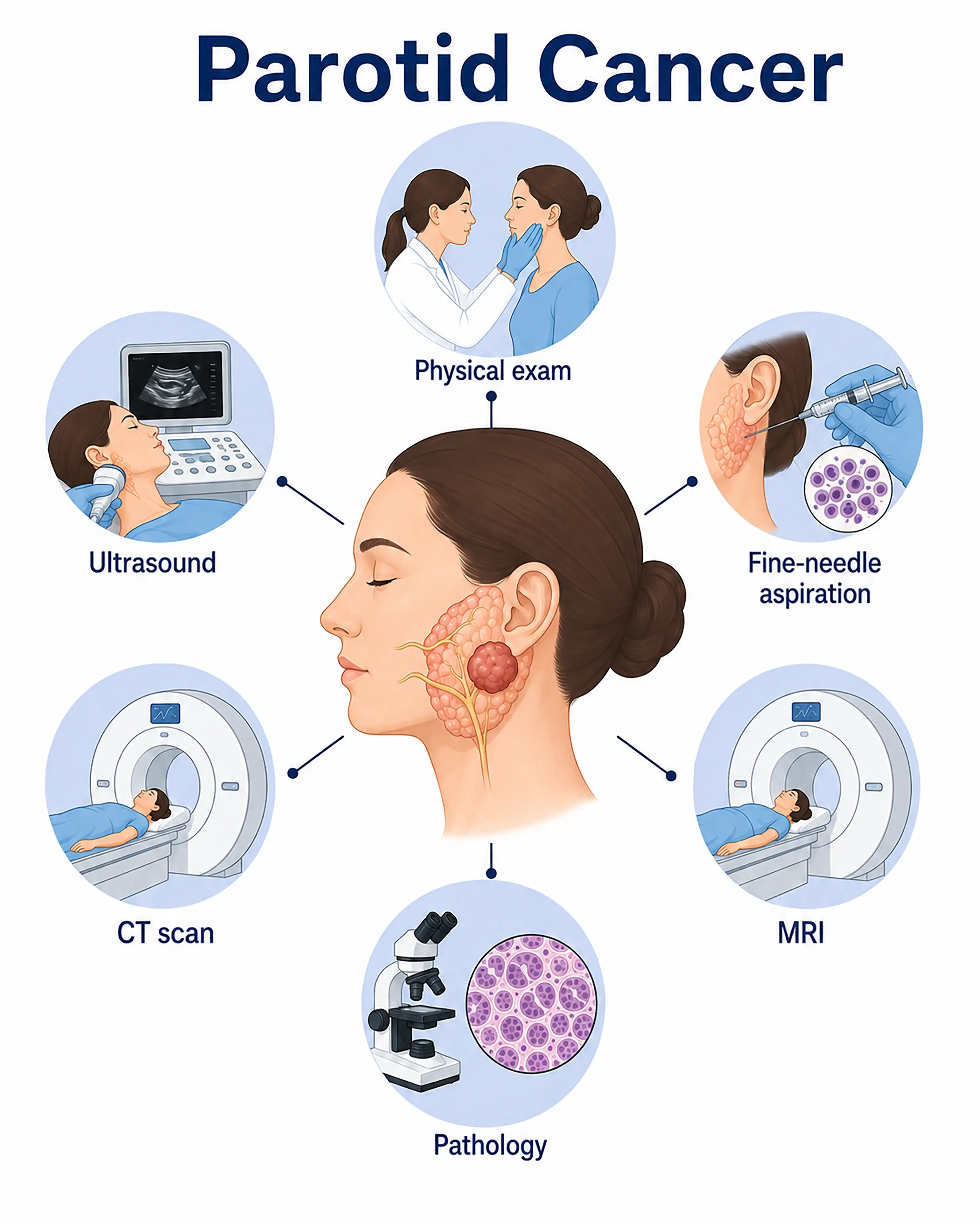
Parotid gland cancer diagnosis by stage
1.Early-stage parotid gland cancer diagnosis
The tumor is confined to the parotid gland, presenting as a painless, hard, poorly mobile lump below the earlobe. Ultrasound shows a hypoechoic mass, and fine-needle aspiration cytology provides an initial benign or malignant assessment.
2.Intermediate-stage parotid gland cancer diagnosis
The tumor enlarges and invades the facial nerve, causing facial numbness or paralysis. Contrast-enhanced CT and MRI show indistinct tumor borders and surrounding tissue involvement. Surgical exploration is needed to establish pathology.
3.Advanced-stage parotid gland cancer diagnosis
The tumor extensively invades the mandible or skull base, or spreads to distant sites. Whole-body PET-CT assesses metastatic extent, and surgery obtains a complete specimen for pathological examination and immunohistochemical typing.
Without timely diagnosis, parotid gland cancer invades the facial nerve, causing permanent facial paralysis and extensive destruction of surrounding tissues. Prognosis after distant metastasis is extremely poor.
When to Seek Medical Attention
Early signs of parotid gland cancer include a painless lump below the earlobe, progressive lump enlargement, facial numbness, or muscle weakness. Consult a doctor through "Chat with a Doctor" for early detection. During an online consultation, the doctor will ask about your symptoms and medical history and guide you toward ultrasound and fine-needle aspiration.
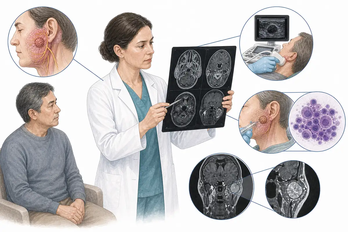
Basis for parotid gland cancer diagnosis
1.Parotid ultrasound
Ultrasound is the preferred noninvasive examination, showing lump size, shape, borders, and internal echoes, providing an initial cystic or solid assessment, and guiding fine-needle aspiration.
2.Fine-needle aspiration biopsy
Ultrasound-guided fine-needle aspiration obtains cytological samples from the lump to initially assess benign or malignant status and pathological type, with an accuracy of 80%-90%.
3.Parotid CT scan
Contrast-enhanced CT clearly shows the tumor's relationship to surrounding tissues, assesses mandibular and skull base invasion, and evaluates cervical lymph node metastasis.
4.Parotid MRI
MRI provides high soft-tissue resolution, showing tumor borders, the course of the facial nerve, and surrounding tissue invasion, supporting preoperative assessment.
5.Open biopsy
Part of the tumor is removed during surgery for rapid frozen-section pathology to establish the diagnosis and guide the surgical plan. This is suitable when fine-needle aspiration results are inconclusive.
6.Facial nerve function assessment
Facial muscle movement is assessed to determine whether the tumor has invaded the facial nerve, providing important information for surgical planning.
7.Cervical lymph node ultrasound
Assesses cervical lymph node metastasis and guides needle biopsy of suspicious nodes to establish tumor stage.
8.Whole-body PET-CT assessment
Whole-body PET-CT assesses distant metastasis and detects small metastatic lesions that conventional imaging may miss, enabling precise staging.
9.Sialography
X-rays are taken after contrast injection into the parotid duct to show filling defects or displacement of the duct system, supporting assessment of tumor extent.
Daily Self-Check
Seek prompt medical care for a painless lump below the earlobe or in front of the ear, especially if it progressively enlarges, hardens, or causes facial numbness. Avoid massaging or applying heat to the lump yourself.
Conclusion
Diagnosis of parotid gland cancer relies on imaging and fine-needle aspiration biopsy. Early detection and characterization of parotid lumps are crucial to preserving facial nerve function and improving cure rates.


