


Osteosarcoma is a tumor formed by the malignant proliferation of mesenchymal cells.Bone tissue consists of osteocytes, bone matrix, and minerals; osteosarcoma is characterized by the direct production of osteoid matrix or tumor bone by the tumor cells and commonly occurs in the metaphyses of long bones.
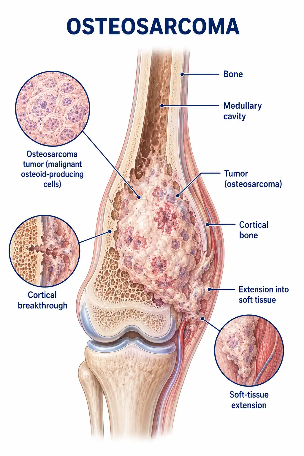
Global overview of osteosarcoma
Osteosarcoma is the most common primary malignant bone tumor, with an incidence of approximately 3 cases per million people annually.It predominantly affects adolescents aged 10–25, with a higher incidence in males than females. The metaphysis of long bones is the most common site of occurrence, with the distal femur and proximal tibia accounting for approximately 60% of cases.
Consult a doctor regarding cancer-related issues
Children and adolescents experiencing symptoms such as persistent bone pain, localized swelling, or unexplained fractures should promptly consult an orthopedic oncologist or an orthopedist.
Main types of osteosarcoma
1. Conventional osteosarcoma
The most common type, accounting for approximately 75% of osteosarcomas; it typically occurs in the metaphysis of long bones and is characterized by the production of tumor bone.
2. Telangiectatic type
Characterized by extensive vascular spaces and hemorrhage, it resembles an aneurysmal bone cyst on imaging but carries a poorer prognosis than the conventional type.
3. Parosteal osteosarcoma
Originates from the bone surface; characterized by slow growth, low malignancy, and a relatively good prognosis; commonly occurs on the posterior aspect of the distal femur.
4. Periosteal osteosarcoma
Originates from the periosteum; its degree of malignancy lies between the conventional and parosteal types; commonly occurs in the diaphysis of long bones.
5. Osteosarcoma of the jaw
Osteosarcoma arising in the jawbones; carries a better prognosis than osteosarcoma of the extremities and is more common in adults.
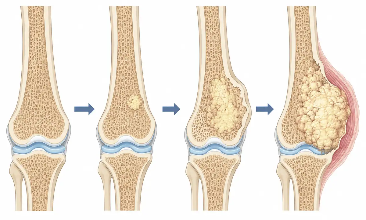
Typical symptoms and signs of osteosarcoma
1. Bone pain
Progressive bone pain is the most common initial symptom; it is notably worse at night, starting as intermittent, vague pain and gradually progressing to constant, severe pain that responds poorly to painkillers.
2. Local swelling
Tumor growth leads to a progressively enlarging mass at the affected site; the mass is hard in texture, the overlying skin temperature is elevated, and varicose veins are visible.
3. Joint dysfunction
When the tumor is located near a joint, it can cause restricted joint movement, pain, and limping, affecting daily activities.
4. Pathological fracture
Tumor destruction of the bone cortex reduces bone strength, allowing fractures to occur under minimal external force; this is known as a pathological fracture.
5. Systemic symptoms
In the advanced stage, patients exhibit systemic symptoms such as weight loss, fatigue, decreased appetite, and fever, indicating disease progression.
6. Pulmonary symptoms
Osteosarcoma is prone to pulmonary metastasis, leading to symptoms such as cough, chest pain, and dyspnea (shortness of breath).
Diagnostic methods for osteosarcoma
1. X-ray examination
X-ray imaging is a key method for the initial screening of osteosarcoma; typical findings include bone destruction, tumor bone formation, and periosteal reactions manifesting as Codman triangles or sunburst patterns.
2. CT scan
Chest CT is used to assess pulmonary metastasis; contrast-enhanced CT can reveal tumor blood supply and the extent of intramedullary infiltration.
3. Magnetic resonance imaging (MRI)
MRI is the optimal method for assessing the extent of tumors within bone and soft tissue, aiding in surgical planning and the evaluation of the feasibility of limb-sparing surgery.
4. Bone scan
Whole-body bone scanning can detect multiple lesions and bone metastases, serving as a valuable reference for staging and treatment planning.
5. Biopsy pathology
Pathological examination of tumor tissue obtained via needle or incisional biopsy is the gold standard for definitive diagnosis and allows for the determination of the pathological type.
6. Laboratory tests
Elevated levels of alkaline phosphatase and lactate dehydrogenase reflect tumor burden and serve as important indicators for prognostic assessment.
Osteosarcoma Treatment Methods
1. Neoadjuvant chemotherapy
Preoperative multi-drug combination chemotherapy is administered to eliminate micrometastases, shrink the tumor to facilitate surgical resection, and assess the tumor's response to chemotherapy.
2. Limb-salvage surgery
Perform wide resection—removing the tumor and surrounding normal tissue—in conjunction with chemotherapy, and reconstruct limb function using prosthetic joints, allografts, or autologous bone grafts.
3. Amputation surgery
Amputation may be required for patients with massive tumors, tumors invading major nerves or blood vessels, or tumors showing a poor response to chemotherapy.
4. Adjuvant chemotherapy
Postoperative chemotherapy is continued to eliminate residual micrometastases and reduce the risk of recurrence; the total treatment course typically lasts 6–12 months.
5. Treatment for pulmonary metastases
For localized pulmonary metastases, surgical resection via thoracotomy may be performed; long-term survival remains possible when combined with chemotherapy.
6. Targeted therapy
Targeted therapies addressing specific molecular markers, such as mTOR inhibitors, are being studied for recurrent or refractory osteosarcoma.
7. Radiotherapy
Osteosarcoma is relatively insensitive to radiotherapy; radiation is primarily used for unresectable lesions or for palliative care, such as pain relief.
8. Rehabilitation therapy
Systematic postoperative rehabilitation training restores limb function and improves quality of life; regular follow-up is required to monitor for recurrence.
Conclusion
Osteosarcoma is the most common primary malignant bone tumor in adolescents.Comprehensive treatment strategies, such as neoadjuvant chemotherapy combined with limb-salvage surgery, have significantly improved patient survival rates and quality of life.


