


Osteosarcoma accounts for approximately 20% of all bone tumors and is the most common primary malignant bone tumor.It predominantly affects adolescents and young adults aged 10–25, with a slightly higher incidence in males than females.It most commonly occurs around the knee joint (distal femur and proximal tibia).With a combination of neoadjuvant chemotherapy and surgery, the five-year survival rate can reach 60%–70%.
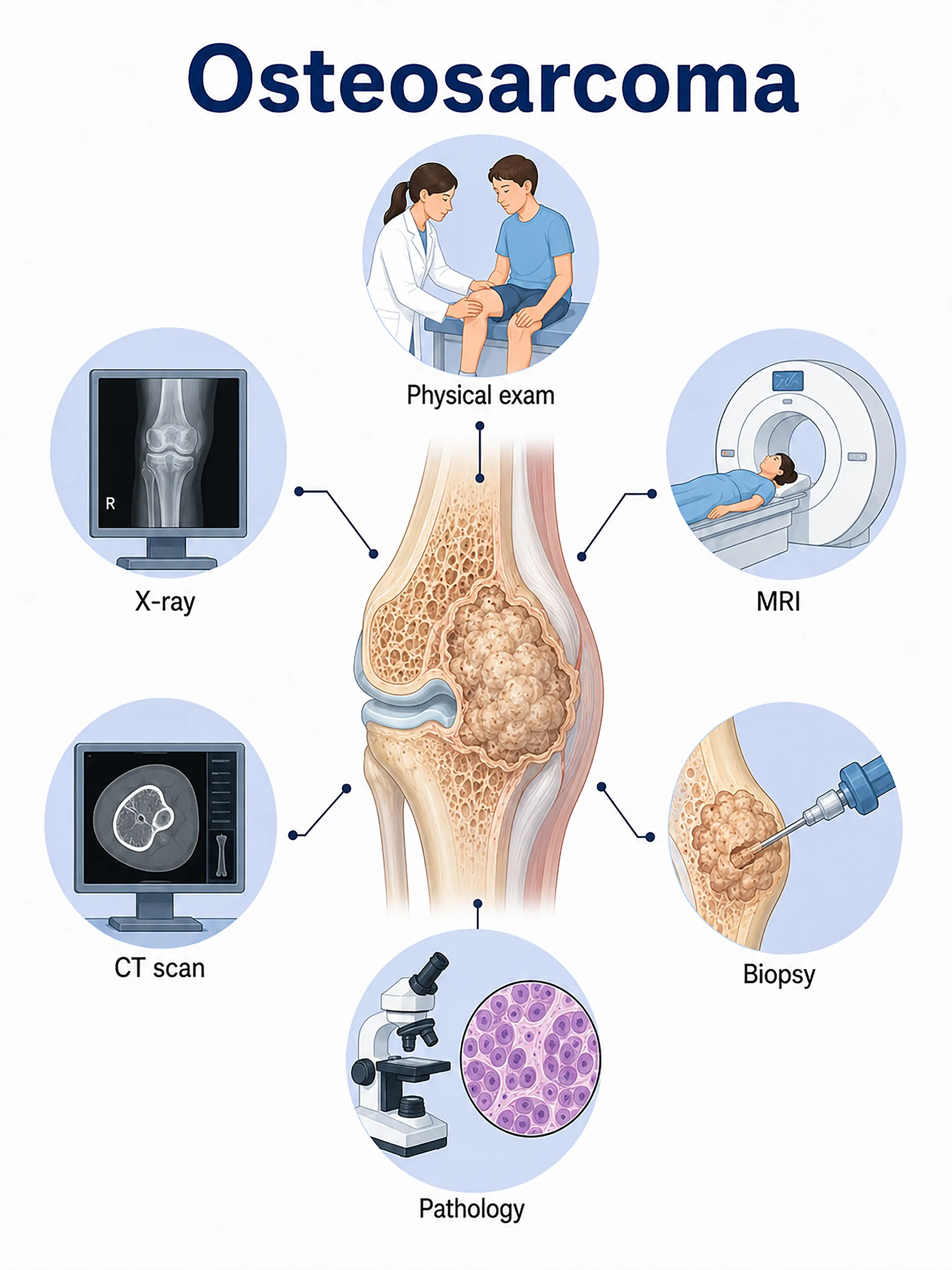
Diagnosis based on osteosarcoma stage
1.Early diagnosis of osteosarcoma
The tumor is confined to the bone and presents with intermittent pain.X-rays show bone destruction and periosteal reactions (Codman triangle, sunburst sign); MRI reveals the extent of intramedullary infiltration; diagnosis is confirmed via needle biopsy.
2.Diagnosis of intermediate-stage osteosarcoma
Tumor breaches the bone cortex, forming a soft-tissue mass.Contrast-enhanced CT and MRI assess tumor extent and adjacent joint involvement; chest CT rules out pulmonary metastasis; biopsy confirms the pathological type.
3.Diagnosis of advanced osteosarcoma
The tumor extensively invades surrounding tissues or metastasizes to the lungs.Whole-body bone scanning and PET-CT are used to assess the extent of metastasis; multiple biopsies are performed to determine the grade, and a comprehensive treatment plan is formulated.
If osteosarcoma is not diagnosed promptly, the tumor progresses rapidly and metastasizes to the lungs; this results in the loss of opportunities for limb-salvage surgery and a significantly poorer prognosis.
When to Seek Medical Attention
Early signs of osteosarcoma include persistent bone pain, localized swelling, and limited mobility.Please consult a doctor via the "Chat with a Doctor" feature to facilitate early detection.During the online consultation, the doctor will ask about the characteristics of your pain and your activity levels, and guide you through the process of obtaining X-ray and MRI examinations.
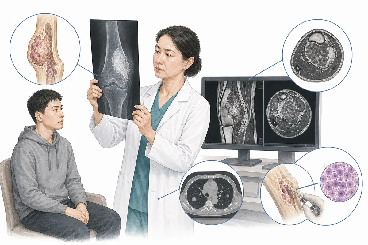
Basis for osteosarcoma diagnosis
1.Plain X-ray examination
X-ray imaging is the preferred initial imaging modality for osteosarcoma; typical findings include bone destruction, periosteal reactions (Codman triangle, sunburst sign), and soft tissue masses.
2.MRI examination
MRI precisely delineates the extent of intramedullary tumor infiltration, the degree of soft-tissue invasion, and skip lesions, making it a core tool for surgical planning and limb-salvage assessment.
3.CT scan
Contrast-enhanced CT is used to assess cortical bone destruction, tumor blood supply, and pulmonary metastasis; chest CT is a routine examination to rule out pulmonary metastasis.
4.Needle biopsy
CT-guided needle biopsy for pathological examination is the gold standard for diagnosing osteosarcoma; the biopsy tract must align with the planned surgical incision.
5.Alkaline phosphatase test
Elevated serum alkaline phosphatase (ALP) indicates active osteogenesis; it is a crucial laboratory marker for osteosarcoma and is used to monitor treatment efficacy and assess prognosis.
6.Whole-body bone scan
Radionuclide bone scanning reveals abnormal tracer uptake at the lesion site while ruling out the possibility of multifocal osteosarcoma or bone metastases.
7.LDH test
Elevated lactate dehydrogenase (LDH) levels indicate a high tumor burden, correlate with prognosis, and serve as an important indicator for supplementary assessment.
8.PET-CT assessment
Whole-body PET-CT assesses distant metastasis and chemotherapy response, offering advantages in detecting metastatic lesions and local recurrence.
9.Preoperative chemotherapy assessment
Following neoadjuvant chemotherapy, MRI is used to assess the tumor necrosis rate (Huvos grade); a necrosis rate >90% is associated with a better prognosis and guides the postoperative chemotherapy regimen.
Daily Self-Check
Adolescents experiencing persistent bone pain—especially pain that worsens at night—or localized swelling should seek medical attention promptly.Avoid mistaking such bone pain for growing pains or sports injuries.
Conclusion
Diagnosis of osteosarcoma relies on X-rays, MRI, and needle biopsy.Early diagnosis is crucial for limb-salvage treatment and improving survival rates.


