


Multiple myeloma is the second most common hematologic malignancy, occurring primarily in middle-aged and elderly individuals.Its main clinical manifestations include bone pain, anemia, renal impairment, and hypercalcemia.Early diagnosis is challenging, and patients often seek medical attention due to bone pain or renal dysfunction.
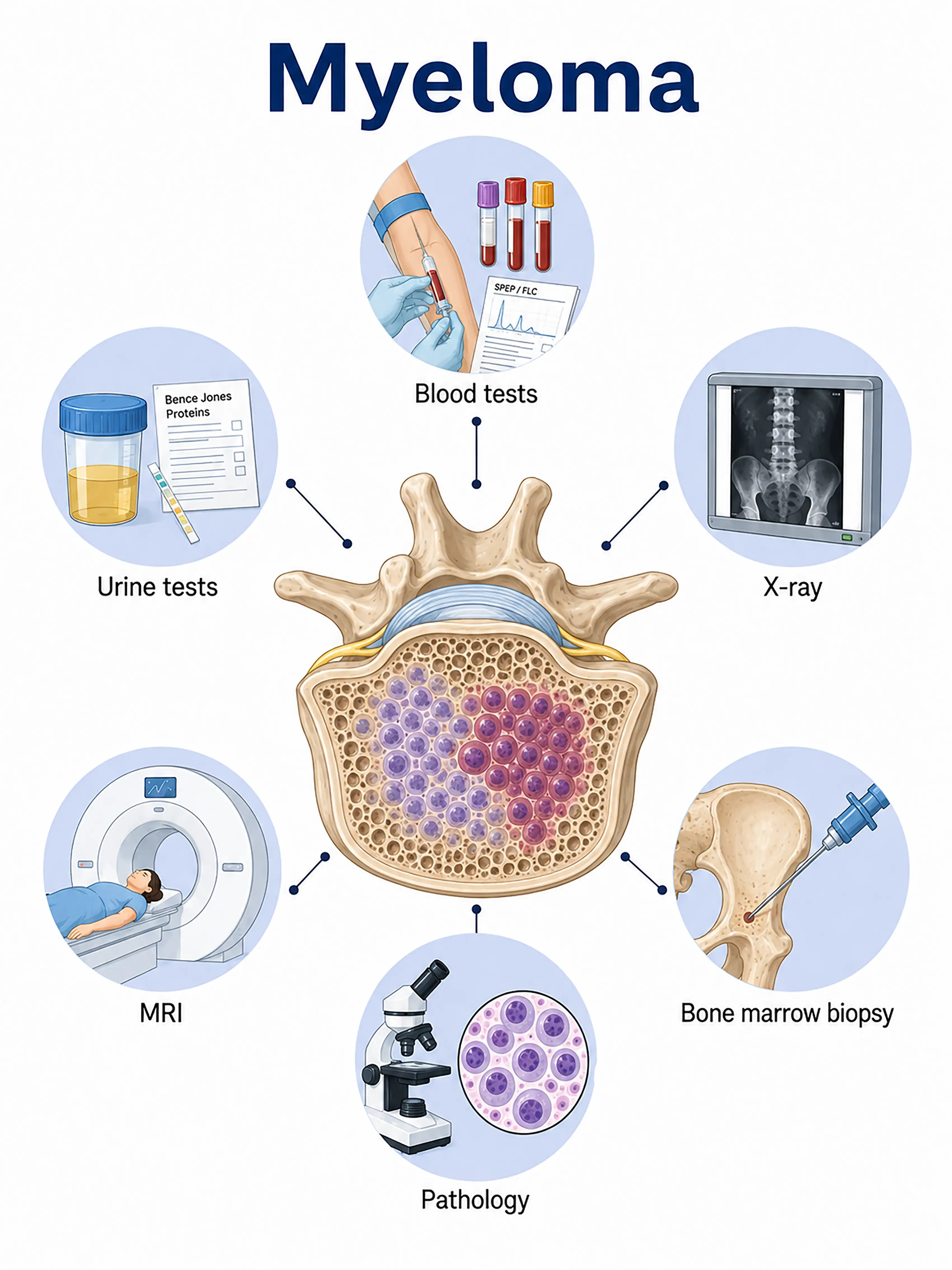
Diagnosis based on myeloma stage
1.Early diagnosis of myeloma
During the asymptomatic phase, an M-protein spike is detected via serum protein electrophoresis.Immunofixation electrophoresis identifies the monoclonal immunoglobulin type, and the free light chain ratio is abnormal; bone marrow aspiration is required for definitive diagnosis.
2.Diagnosis of intermediate-stage myeloma
Presentation of typical CRAB symptoms.Bone marrow aspiration shows >10% plasma cells; serum protein electrophoresis reveals M-protein; X-ray or MRI shows osteolytic lesions;diagnosis is confirmed by biopsy.
3.Diagnosis of advanced myeloma
Extensive bone destruction with multiple pathological fractures; renal failure.Whole-body MRI or PET-CT assesses extramedullary lesions; serial biopsies monitor clonal evolution.
If myeloma is not diagnosed in time, the tumor can lead to extensive bone destruction, renal failure, and recurrent infections, severely affecting quality of life and survival.
When to Seek Medical Attention
Early signs of myeloma include persistent bone pain, anemia and fatigue, abnormal renal function, and manifestations of hypercalcemia.Please consult a doctor via the "Chat with a Doctor" feature for early detection.During the online consultation, the doctor will ask about your symptoms and laboratory test results, and guide you through tests such as serum protein electrophoresis and bone marrow examination.

Diagnostic Criteria for Myeloma
1.Serum protein electrophoresis
Serum protein electrophoresis can detect an M-protein (monoclonal immunoglobulin) peak and is the preferred laboratory test for screening and diagnosing multiple myeloma.
2.Immunofixation electrophoresis
Immunofixation electrophoresis identifies the heavy and light chain types of M-protein (IgG, IgA, IgD, or light chain type) and is a crucial method for definitive diagnosis and subtyping.
3.Bone marrow aspiration and biopsy
Bone marrow aspiration smears and biopsies showing an increased proportion of plasma cells (>10%) accompanied by morphological abnormalities constitute the gold standard for diagnosing myeloma.
4.Free light chain assay
Measure serum concentrations and the ratio of free kappa and lambda light chains; an abnormal ratio supports the diagnosis of light-chain myeloma and offers high sensitivity.
5.Whole-body MRI
Whole-body MRI (WB-MRI) assesses bone marrow infiltration and bone destruction; it can detect early lesions invisible on X-rays and is the preferred method for evaluating bone disease.
6.Skeletal X-ray examination
A whole-body skeletal X-ray examination reveals osteolytic lesions, osteoporosis, and pathological fractures; it is a traditional method for assessing bone damage.
7.PET-CT assessment
Whole-body PET-CT assesses extramedullary lesions, metabolic activity, and treatment response, providing valuable information for prognosis.
8.Renal function and electrolytes
Measure levels of serum creatinine, blood urea nitrogen (BUN), calcium, and phosphorus to assess the degree of renal impairment and hypercalcemia.
9.Flow cytometry
Flow cytometry analysis of bone marrow specimens detects abnormal plasma cell immunophenotypes, aiding in diagnosis and monitoring for minimal residual disease.
Daily Self-Check
Seek medical attention promptly if unexplained bone pain, anemia, or abnormal renal function occurs.High-risk individuals over the age of 40 should undergo regular screening via serum protein electrophoresis.
Conclusion
Diagnosis of myeloma relies on serum protein electrophoresis, immunofixation electrophoresis, and bone marrow aspiration.Early diagnosis can significantly reduce complications and improve survival.


