


What is melanoma
Melanoma is a tumor formed by the malignant proliferation of melanocytes.Melanocytes are distributed in areas such as the basal layer of the skin, the eyes, and mucous membranes, producing melanin to protect the skin from ultraviolet (UV) damage.
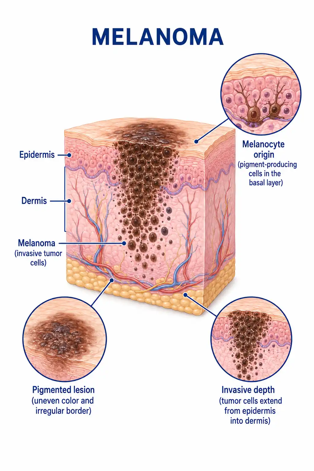
Global status of melanoma
Melanoma incidence is high among Caucasians, and rates in my country have also been rising in recent years.It predominantly affects adults and can occur on the skin, in the eyes, or on mucous membranes, with cutaneous (skin) melanoma being the most common form.
Consult a doctor regarding cancer-related issues
Patients noticing changes in the shape, color, or size of a pigmented nevus, or the appearance of a new irregular pigmented lesion, should promptly consult a dermatologist or oncologist.
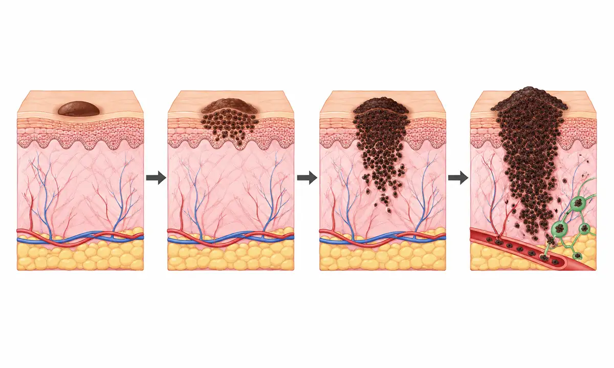
Main types of melanoma
1. Superficial spreading type
The most common type of cutaneous melanoma; it features a prolonged horizontal growth phase, typically occurs on the trunk and limbs, and has a prognosis closely related to the depth of invasion.
2. Nodular type
Characterized primarily by vertical growth, it infiltrates the deep dermis at an early stage, grows rapidly, and carries a poor prognosis.
3. Lentigo maligna type
It typically occurs in the head and neck region of elderly patients, grows slowly, and undergoes a prolonged phase of in situ growth before progressing to vertical invasion.
4. Acral lentiginous type
It typically occurs on the palms, soles, and beneath the nails; it is the most common subtype among Asians and is easily overlooked.
5. Mucosal melanoma
Occurs on the mucosa of the oral cavity, nasal cavity, gastrointestinal tract, and genitourinary tract; the prognosis is generally poor.
Typical symptoms and signs of melanoma
1. Changes in pigmented nevi
Changes in an existing pigmented nevus—such as asymmetry, irregular borders, uneven color, or increased diameter—are important warning signs of melanoma.
2. New pigmented lesion
Pigmented skin lesions appearing in adulthood, particularly those that are rapidly growing, uneven in color, or have irregular borders.
3. Ulcer bleeding
Ulceration, bleeding, or crusting on the tumor surface indicates deep tumor infiltration.
4. Itching and pain
Sensory abnormalities such as itching, pain, or tenderness in a pigmented nevus.
5. Satellite lesion
The appearance of small pigmented satellite lesions around the primary tumor indicates local tumor dissemination.
6. Lymph node enlargement
Enlarged regional lymph nodes that are hard in texture and have poor mobility suggest possible lymph node metastasis.
How is melanoma diagnosed
1. Dermoscopy
Dermoscopy allows for the magnified observation of the fine structures of pigmented lesions, aiding in the differentiation between benign and malignant conditions and guiding biopsies.
2. Biopsy pathology
Complete excision of the suspicious lesion followed by pathological examination is the gold standard for diagnosis; parameters such as Breslow thickness, ulceration status, and surgical margin status must be determined.
3. Sentinel lymph node biopsy
For melanomas thicker than 1 mm, sentinel lymph node biopsy is an important method for assessing lymph node metastasis status.
4. Imaging examination
CT, MRI, and PET-CT are used for staging and assessing distant metastases, including those to the lungs, liver, bones, and brain.
5. Genetic testing
Test for gene mutations such as BRAF, NRAS, and KIT to guide targeted therapy and prognostic assessment.
6. LDH test
Elevated lactate dehydrogenase (LDH) levels indicate a high tumor burden and serve as a significant prognostic indicator for stage IV melanoma.
What are the treatment methods for melanoma
1. Wide resection
The standard treatment for early-stage melanoma involves wide local excision; the extent of resection is determined by Breslow thickness to ensure adequate safety margins.
2. Lymph node dissection
Regional lymph node dissection is required in cases of positive sentinel lymph nodes or clinical lymph node metastasis.
3. Targeted therapy
The combination of BRAF inhibitors and MEK inhibitors is used for BRAF-mutated advanced melanoma and can significantly prolong survival.
4. Immunotherapy
PD-1 inhibitors and CTLA-4 inhibitors are important therapeutic options for advanced melanoma, enabling long-term survival in some patients.
5. Adjuvant immunotherapy
Adjuvant therapy with PD-1 inhibitors in high-risk patients after surgery can reduce the risk of recurrence.
6. Adjuvant targeted therapy
High-risk patients with BRAF mutations receive targeted drug therapy as an adjuvant treatment following surgery.
7. Isolated limb perfusion
For local treatment of multiple cutaneous metastases on the limbs; local lesions are controlled via hyperthermic perfusion with chemotherapeutic agents.
8. Follow-up monitoring
Postoperative follow-up involving regular skin examinations and imaging is crucial to monitor for recurrence and metastasis.
Conclusion
While melanoma is a less common type of skin cancer, it is highly malignant.Patient prognosis has improved significantly through early detection, standardized surgery, and immunotherapy or targeted therapy.


