


The global incidence of melanoma is rising, with the highest rates observed in Caucasian populations. Ultraviolet (UV) exposure is a primary risk factor;common sites of occurrence include the back, legs, and head and neck.
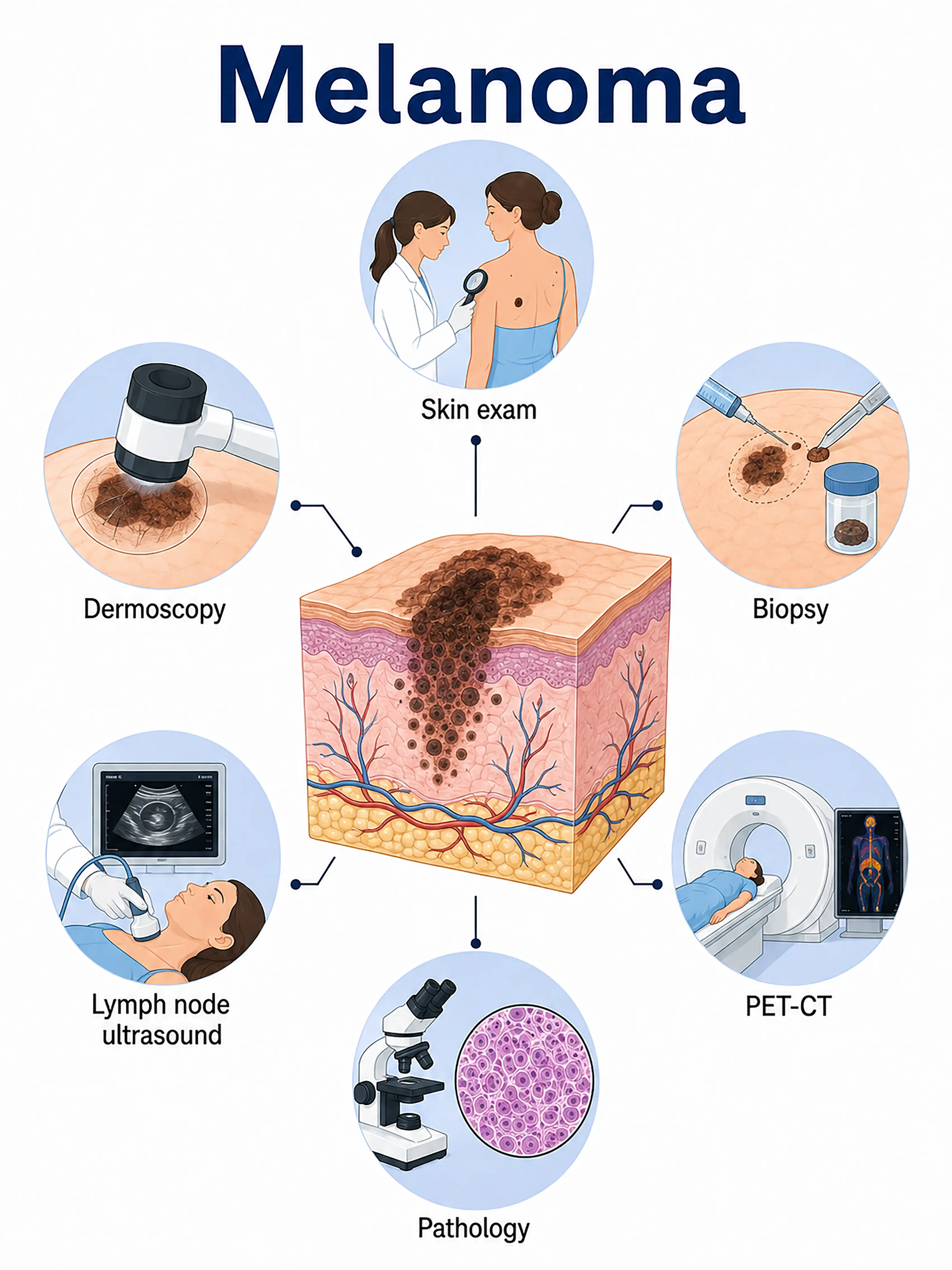
Staging and diagnosis of melanoma
Early symptoms of melanoma
Early signs include changes in the color, shape, or size of an existing mole, as well as irregular borders and uneven coloration.Itching, bleeding, or crusting may occur, and the diameter is often greater than 6 mm.
Symptoms of intermediate-stage melanoma
In the intermediate stage, the lesion enlarges and thickens significantly;the surface may ulcerate or develop nodules, and the color deepens or becomes variegated.Regional lymph nodes may become enlarged, feeling hard and fixed upon palpation.
Symptoms of advanced melanoma
Advanced-stage tumors extensively infiltrate subcutaneous tissues and deep structures, resulting in satellite lesions and distant metastases.Metastasis to the liver, lungs, bones, and brain leads to corresponding organ dysfunction and systemic failure.
Consequences of failing to receive a timely diagnosis in the early stage
Failure to receive a timely diagnosis in the early stage leads to increased tumor thickness and a significantly higher risk of lymph node and distant metastasis.Patients diagnosed at an advanced stage have limited treatment options and significantly lower survival rates.
When to Seek Medical Attention
If a skin mole shows changes in color, irregular borders, increased diameter, elevation, or ulceration, visit a dermatologist immediately for a dermoscopy examination as soon as possible.
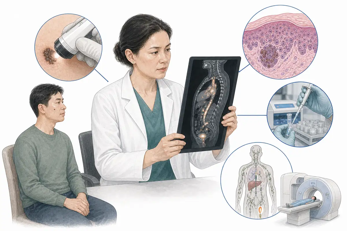
Basis for Melanoma Diagnosis
1. Dermoscopy assessment
Dermoscopy allows for the magnified observation of pigment patterns, vascular structures, and pigment distribution; it is an important tool for early screening and distinguishing between benign nevi and melanoma.
2. Pathological tissue biopsy
Complete excision of the lesion for pathological examination—to confirm the diagnosis of melanoma and measure tumor thickness and depth of invasion—is the gold standard for diagnosis.
3. Sentinel lymph node biopsy
Sentinel lymph node biopsy is performed for melanomas thicker than 0.8 mm to assess lymph node metastasis status and guide staging and the extent of surgery.
4. Whole-body imaging
Chest/abdominal CT and PET-CT are used to assess distant metastasis to the lungs, liver, bones, and brain, determine tumor stage, and guide subsequent treatment plans.
5. Tumor marker testing
Elevated serum LDH levels indicate disease progression and metastasis; they serve as a prognostic indicator for stage IV melanoma and are used for dynamic monitoring of treatment efficacy.
6. Genetic test analysis
Test for gene mutations such as BRAF, NRAS, and c-KIT to guide the selection of targeted therapies, assess prognosis, and formulate personalized treatment plans.
7. Wide excision of the primary tumor
Excision of the lesion along with a surrounding margin of normal skin and subcutaneous tissue; the surgical margins are sent for pathological examination to ensure they are negative, thereby reducing the risk of recurrence.
Daily Self-Check
Regularly observe skin moles for changes using the ABCDE rule (Asymmetry, Border, Color, Diameter, Evolution); seek medical attention promptly if abnormalities are detected.
Conclusion
Early recognition of melanoma is crucial;pay attention to changes in skin moles, perform regular self-examinations, and seek medical attention promptly if abnormalities are found, as early diagnosis significantly improves prognosis.


