


Medullary Thyroid Cancer Diagnosis
Medullary thyroid cancer accounts for 1%-2% of thyroid cancers and is classified as sporadic or familial (MEN2 syndrome). It usually occurs at ages 40-60. Familial disease is associated with RET proto-oncogene mutations and allows genetic screening and preventive thyroidectomy.
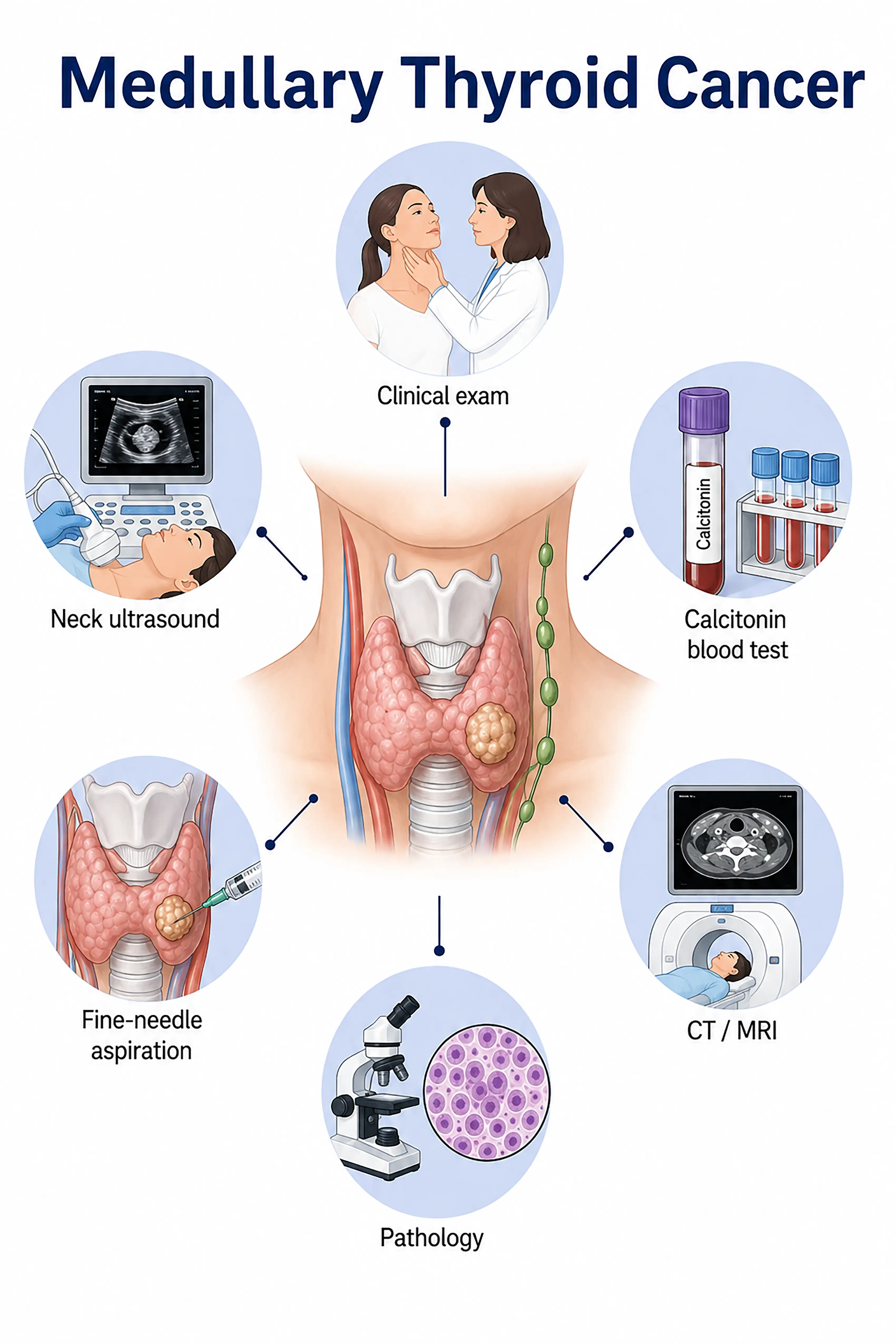
Medullary thyroid cancer diagnosis by stage
1 Early-stage medullary thyroid cancer diagnosis
Palpation reveals a thyroid nodule and serum calcitonin is elevated (>100 pg/mL is highly suggestive). Ultrasound-guided fine-needle aspiration with washout calcitonin testing supports confirmation.
2 Intermediate-stage medullary thyroid cancer diagnosis
Calcitonin and CEA rise markedly, with enlarged cervical lymph nodes. Ultrasound and CT assess tumor extent and nodal metastasis, while RET testing determines whether the disease is familial.
3 Advanced-stage medullary thyroid cancer diagnosis
The tumor invades surrounding tissue or spreads to the liver, lungs, and bones. Whole-body imaging assesses metastatic extent, while genetic testing guides targeted therapy such as selpercatinib.
Without timely diagnosis, medullary thyroid cancer invades surrounding tissues and metastasizes, with persistently elevated calcitonin and CEA and a poor prognosis.
When to Seek Medical Attention
Early signs include neck lumps, diarrhea, and facial flushing. Consult a doctor through "Chat with a Doctor" for early detection. During an online consultation, the doctor will ask about your family history and symptoms and guide you toward calcitonin testing and genetic screening.
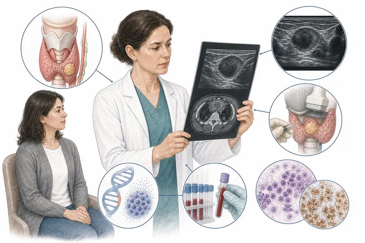
Basis for medullary thyroid cancer diagnosis
1 Serum calcitonin testing
Calcitonin is a specific marker. Serum levels >100 pg/mL strongly support medullary carcinoma and are central to screening and monitoring.
2 Serum CEA testing
Carcinoembryonic antigen (CEA) is often elevated. Combined testing with calcitonin improves diagnostic accuracy and is important for postoperative monitoring.
3 Fine-needle aspiration biopsy
Ultrasound-guided fine-needle aspiration with calcitonin measurement in the washout fluid can accurately confirm or exclude medullary carcinoma, especially when serum calcitonin is inconclusive.
4 Thyroid ultrasound
Ultrasound assesses thyroid nodule size, shape, borders, and calcification, guides fine-needle aspiration biopsy, and monitors cervical lymph node metastasis.
5 RET genetic testing
RET proto-oncogene mutations such as M918T and C634R are tested to identify familial medullary carcinoma (MEN2A or MEN2B) and guide family screening.
6 Neck CT and MRI
Contrast-enhanced CT and MRI assess local invasion and cervical lymph node metastasis to inform surgical planning.
7 Whole-body imaging assessment
CT, MRI, and PET-CT assess distant spread to the liver, lungs, bones, and other sites and are recommended for complex disease or persistently elevated calcitonin.
8 Preoperative calcitonin doubling time
Calcitonin and CEA doubling times (DT) are important parameters for assessing progression rate and prognosis and guide treatment decisions.
9 Immunohistochemistry testing
Immunohistochemical analysis of pathology specimens tests neuroendocrine markers such as calcitonin, CEA, and chromogranin A to support diagnosis.
Daily Self-Check
People with a family history should have regular calcitonin testing and RET screening. A neck lump with diarrhea or facial flushing warrants prompt care.
Conclusion
Diagnosis relies on calcitonin testing and fine-needle aspiration biopsy. RET testing is crucial for familial screening. Early total thyroidectomy offers the best chance of cure.


