


Laryngeal cancer accounts for a significant proportion of head and neck malignancies. It is markedly more common in men, with a male-to-female ratio of about 8-10:1. Smoking and alcohol consumption are the main risk factors, and it usually occurs at ages 50-70. It is classified by location as supraglottic, glottic, or subglottic.
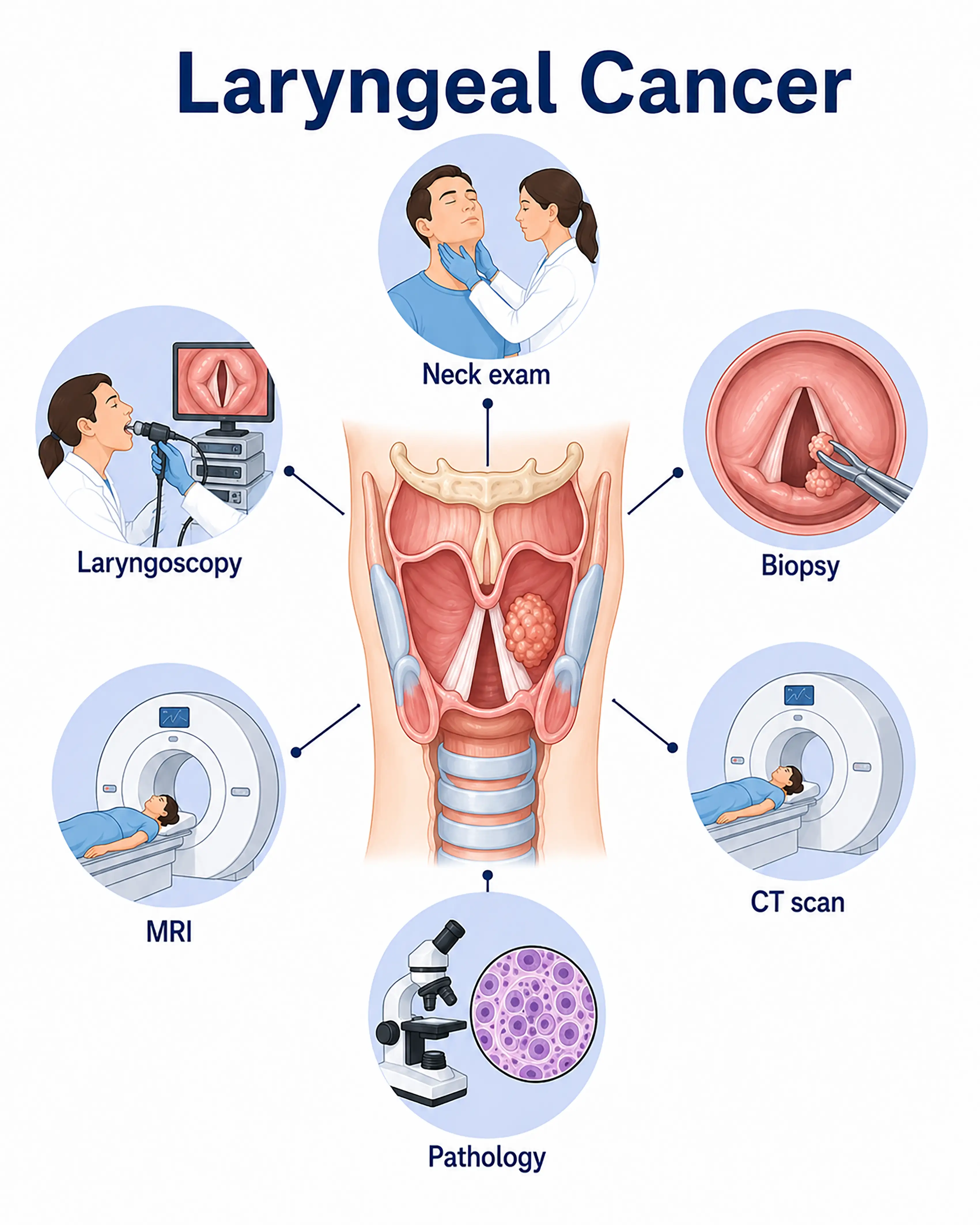
Laryngeal cancer diagnosis by stage
1.Early-stage laryngeal cancer diagnosis
Glottic cancer presents with persistent hoarseness and polypoid or papillary growths on the vocal cords. Laryngoscopy identifies localized vocal cord lesions, and biopsy establishes the pathological diagnosis.
2.Intermediate-stage laryngeal cancer diagnosis
The tumor invades structures adjacent to the vocal cords or the supraglottis. Laryngoscopy reveals ulcerative or cauliflower-like masses, while contrast-enhanced CT assesses cartilage invasion and cervical lymph node metastasis.
3.Advanced-stage laryngeal cancer diagnosis
The tumor extensively invades beyond the larynx or spreads to distant sites. Whole-body PET-CT assesses lung and bone metastases, surgical exploration establishes the stage, and pathology guides chemoradiotherapy planning.
Without timely diagnosis, laryngeal cancer invades laryngeal cartilage and surrounding tissues and has a higher risk of distant metastasis. Total laryngectomy may become necessary, causing permanent loss of natural voice.
When to Seek Medical Attention
Early signs of laryngeal cancer include persistent hoarseness lasting more than two weeks, a foreign body sensation in the throat, painful swallowing, and blood-stained sputum. Consult a doctor through "Chat with a Doctor" for early detection. During an online consultation, the doctor will ask about your smoking and drinking history and symptoms and guide you toward laryngoscopy.
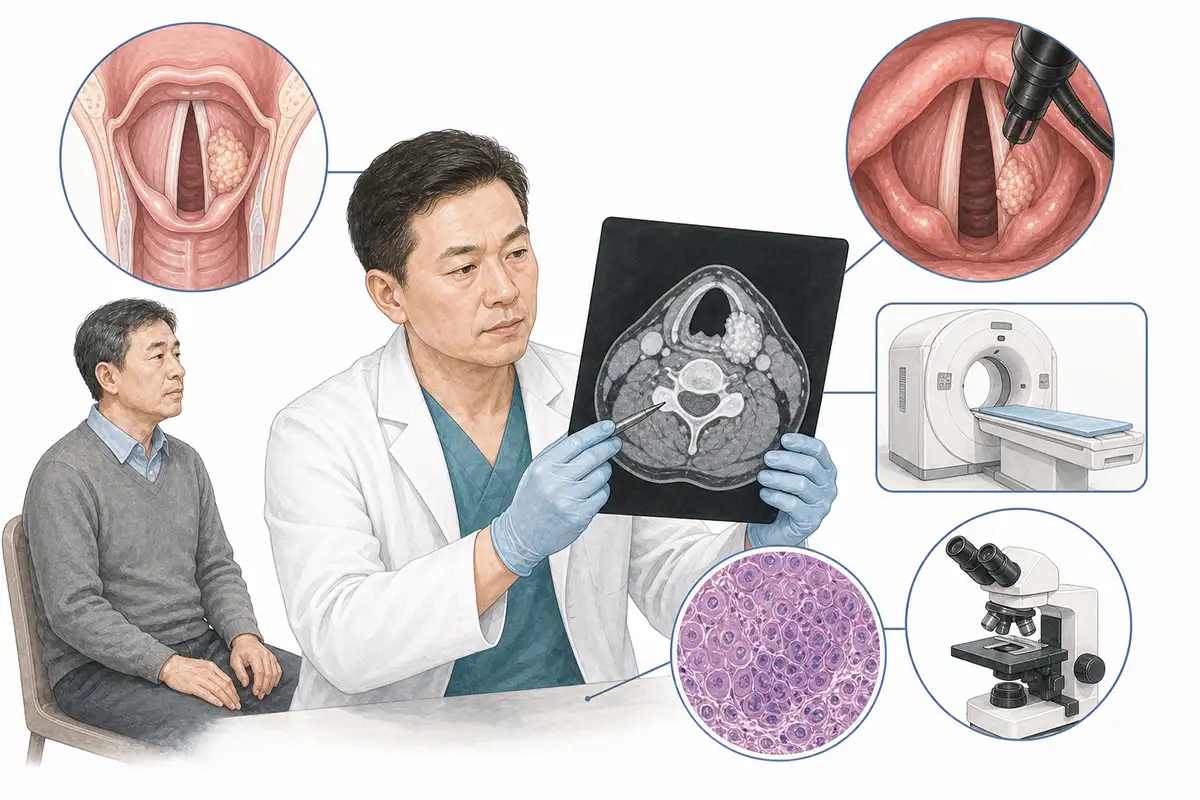
Basis for laryngeal cancer diagnosis
1.Indirect laryngoscopy
A handheld laryngeal mirror examines the throat and vocal cords to identify polypoid or papillary vocal cord growths, providing a simple initial screening method.
2.Electronic laryngoscopy
High-definition magnification enables electronic laryngoscopy to clearly show subtle laryngeal mucosal lesions, vocal cord movement, and tumor extent.
3.Histopathological biopsy
Lesion tissue is sampled with forceps under laryngoscopy or suspension laryngoscopy for pathological examination, the gold standard for diagnosing laryngeal cancer. Squamous cell carcinoma accounts for more than 95%.
4.Neck CT scan
Contrast-enhanced CT assesses invasion of laryngeal cartilage and the pre-epiglottic space and cervical lymph node metastasis, which is crucial for staging and surgical planning.
5.Neck MRI
MRI provides high soft-tissue resolution, assessing the tumor's relationship to surrounding muscles, blood vessels, and nerves and distinguishing tumor from edema.
6.Laryngeal ultrasound
High-frequency ultrasound assesses vocal cord lesions and cervical lymph node metastasis and guides lymph node needle biopsy.
7.Whole-body PET-CT assessment
Whole-body PET-CT assesses distant metastasis and detects small metastatic lesions that conventional imaging may miss, enabling precise staging.
8.Voice function assessment
A voice analyzer assesses vocal cord vibration and voice quality, providing a reference for surgical planning and postoperative rehabilitation.
9.Laryngeal electromyography
Assesses recurrent laryngeal nerve function, distinguishes causes of vocal cord paralysis, and determines whether the tumor has invaded nerves.
Daily Self-Check
Seek immediate medical care for persistent hoarseness lasting more than two weeks, a foreign body sensation in the throat, painful swallowing, or blood-stained sputum. Quitting smoking and limiting alcohol are important preventive measures.
Conclusion
Early diagnosis of laryngeal cancer is crucial for preserving laryngeal function and improving survival. Persistent hoarseness is an important warning sign of glottic cancer and warrants prompt laryngoscopy.


